

INTRODUCTION
Subcutaneous tissue inoculation in murine preclinical model, like lymphoma model, is an efficient and cost-effective way to evaluate in vivo the response of tumors to novel therapies. The tumoral cells are implanted into the flank of a mice. Tumor is then usually assessed using a caliper, the current gold standard method to monitor the volume growth. The caliper has however some limitations. It cannot distinguish the tumor itself from the surrounding inflammatory or fibrotic tissue. It is also an invasive method that measures only the external surface of a tumor ignoring larger and deeper lesion. On contrary, MRI is a noninvasive approach that allows (1) a 3D assessment of the tumoral volume (2) and in vivo monitoring of lesional growth. This project aims at showing that MRI provides better accuracy and reliability compared to caliper measurements for assessing subcutaneous lymphoma model, supporting its broader application in large-scale preclinical oncology studies.
METHODS
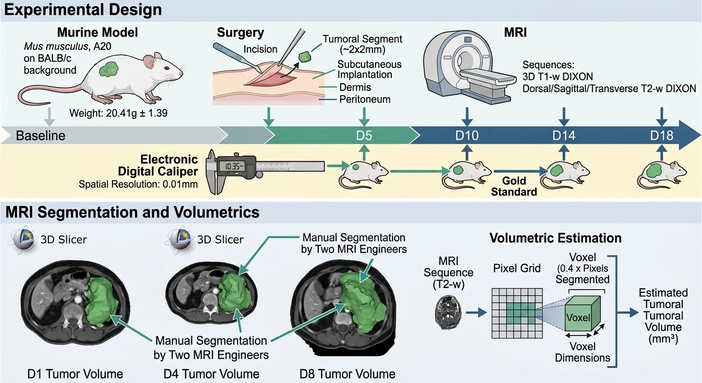
Experimental Design
• Model: Female murine model (Mus musculus, A20 on a BALB/c background; weight=20.41g±1.39)
• Sample Size: 10 mice
• Surgery: Subcutaneous implantation of approx. 2x2mm of tumoral segment from B lymphoma between dermis and peritoneum
• MRI: 1.5T MRI (Signal GE Explorer, General Electrics, Chicago, Illinois, USA)
• Sequences: 3D T1-weighted imaging DIXON; Dorsal, Sagittal and Transverse T2-weighted imaging DIXON
• Imaging: MRI data acquisition was performed at 5 time points (Baseline, D5, D10, D14 and D18) to monitor changes in tumoral volume • Gold Standard: Electronic Digital Caliper (e.g., Spatial Resolution = 0.01mm)
MRI Segmentation and Volumetrics
Each lymphoma model volume at D1, D4 and D8 were manually segmented by two MRI application engineers on the T2-weighted MRI sequence, using 3D Slicer. This approach was privileged as automated algorithms do not perform well due to the irregular nature of tumor shape. The tumoral volumes were finally estimated by multiplying the number of pixels segmented by the
dimensions of the voxel (0.4 x 0.4mm) in the image.
RESULTS
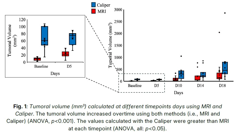
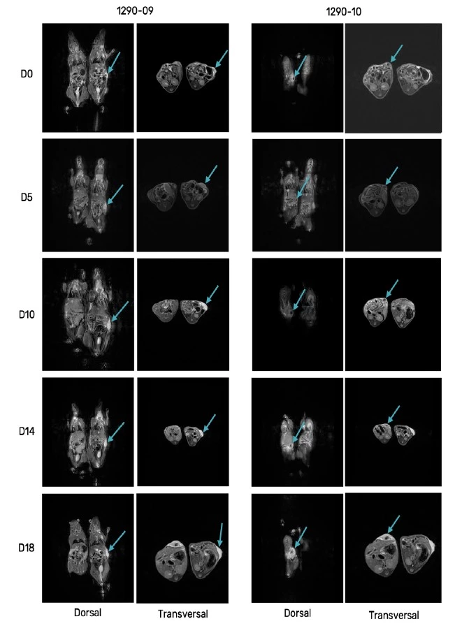
Differences in the lymphoma model volumetrics were observed between the Caliper and MRI. Several reasons can explain these results. First, Caliper offers an approximation of the tumoral volume (i.e., sphere). Second, the visible mass measured with Caliper included all tissues directly surrounding the lymphoma model. On the contrary, MRI was able to estimate an accurate 3D volume of the tumor. This imaging modality was also able to distinguish the tumoral mass from the surrounding tissues (i.e., fat, inflammation and dysplastic content) at each timepoint until the end of the study (D18) using a T2-weighted imaging DIXON sequence. This result supports the use of MRI to distinguish the tumoral mass from the surrounding oedema/inflammation.
RELEVANCE TO CANCER RESEARCH
In this second white paper realised with Hawkcell, we demonstrate that MRI is a powerful, non-invasive imaging modality that can play a crucial role in cancer research by enabling precise and reproducible in vivo measurement of tumoral volume. This is particularly valuable in early-phase drug development, where accurate, longitudinal monitoring of tumor growth or regression is essential for assessing the efficacy of novel therapeutic agents. MRI can capture detailed anatomical and functional information, including tumor vascularity, necrosis, and diffusion characteristics, provides a multidimensional view of tumor biology. It also offers high soft tissue contrast, which is particularly beneficial for visualizing tumors in anatomically complex regions (e.g., brain, liver, or pancreas).
MRI allows researchers to monitor in vivo tumor progression over time within the same animal. This not only enhances the statistical power of the preclinical studies but also aligns with the 3Rs by minimizing the number of animals needed. MRI datasets can also be retrospectively post-processed, allowing researchers to extract new insights (e.g., texture analysis, volumetric changes, or radiomics) even after the completion of the study. This adds significant value and flexibility to data interpretation, supporting both hypothesis-driven and exploratory analyses.
CONCLUSION
MRI and the associated post-processing methods to measure tumoral volume is a more sensitive, reliable and reproducible approach to assess the tumoral volume in longitudinal studies compared to the currently gold standard, the Caliper. MRI also lowers the need for additional animal cohorts for intermediate euthanasia, by enabling precise, non-invasive monitoring of tumoral progression over time. These not only enhance study efficiency and data quality but also aligns with the principles of the 3Rs in animal research.
By integrating MRI into cancer research workflows, scientists gain a robust, scalable, and ethically responsible tool to evaluate tumor response, monitor disease progression, and support the translation of preclinical findings into clinical trials. Given its accuracy, longitudinal capability, and ethical advantages, MRI should be considered the new standard for preclinical oncology studies.
This white paper provides a foundation for further studies that can refine the experimental design, optimize the treatment regimen, and expand on the use of MRI in oncology preclinical research.
The MRI imaging is a very good complementary technique to luciferase bioluminescent imaging of live animal.
Contact our Team to learn more
Download the new white paper here
FAQ
Subcutaneous tissue inoculation in a murine preclinical setting provides a cost-effective method for cancer research. In this process, cells of a lymphoma model are implanted into the flank of female mice. Response to new therapeutic compounds is subsequently monitored in vivo. Traditional evaluations are performed with calipers to track outer dimensions over time. However, deeper lesions are often ignored by surface measurements. Unintended inclusion of adjacent inflammatory or fibrotic tissue is also caused by manual measurement methods. Consequently, improved techniques are required so that underlying lesion expansion is captured without invasive physical contact.
Female mice of the species Mus musculus with an A20 on a BALB/c background and a mean weight of 20.41g±1.39 are selected for testing. A small tumoral segment measuring approximately 2x2mm from B lymphoma is subcutaneously implanted. Placement is surgically completed between the dermis and the peritoneum across a sample size of 10 mice. Longitudinal changes in tissue structure are then documented across five pre-determined intervals. Observations are conducted at Baseline, D5, D10, D14, and D18. Through this structured schedule, comparative data regarding cellular expansion is collected under controlled laboratory conditions.
Scanning procedures are performed using a 1.5T MRI system identified as Signal GE Explorer from General Electrics, Chicago, Illinois, USA. Specific imaging protocols are configured to acquire detailed anatomical information. Three-dimensional T1-weighted imaging DIXON sequences are applied during the scanning sessions. Additionally, Dorsal, Sagittal, and Transverse T2-weighted imaging DIXON sequences are utilized for internal visualization. Digital information collected from these scans is saved for subsequent volumetric processing. High spatial resolution images are thus gathered without causing physical disruption to the animal.
An Electronic Digital Caliper with a spatial resolution of 0.01mm is employed as a comparative gold standard. Dimensional values are gathered manually from the exterior boundaries of the growth. Physical dimensions are recorded at specified intervals to estimate overall volume expansion. However, external boundary tracking cannot separate neoplastic cells from adjacent non-cancerous swelling. Inaccurate calculations may occur when inner necrotic zones or deep tissue extensions are present. Therefore, manual measurements are combined with non-invasive imaging techniques to verify observational consistency across all subjects.
Volumetric measurements for each lymphoma model at D1, D4, and D8 are derived from T2-weighted MRI sequences. Image segmentation is manually executed by two trained MRI application engineers. Outlines of the target mass are delineated slice by slice to construct a full three-dimensional representation. Tissue boundaries are identified based on signal contrast variations inherent to the acquired sequence. Internal lesion structures are isolated from non-tumoral surrounding regions during this process. Independent evaluations by multiple specialists ensure that boundary selection remains consistent throughout the longitudinal dataset.
Volumetric assessment in three dimensions is directly enabled through magnetic resonance protocols. Unlike physical calipers, deep-seated lesions and internal structural variations are rendered clearly by magnetic signals. Surrounding inflammatory reactions or fibrotic tissues are effectively differentiated from actual tumoral boundaries. Furthermore, non-invasive imaging allows continuous monitoring of the same animal over extended timeframes. Animal stress is reduced while data quality from every session is improved. Consequently, superior accuracy is attained when treatment efficacy is evaluated in preclinical studies.
Data collection across the study is planned at five discrete time points. Baseline scans are performed prior to tracking subsequent growth patterns in the subjects. Follow-up imaging sessions are conducted on D5, D10, D14, and D18. Through this regular timetable, continuous monitoring of lesional growth is achieved in vivo. Tumour progression across multiple weeks is measured without requiring terminal procedures at each step. Longitudinal tracking across identical subjects yields reliable volumetric curves for statistical analysis.
Repeated non-invasive measurements are permitted when magnetic resonance tools are integrated into research protocols. Because individual subjects are imaged continuously from Baseline to D18, fewer animals are required per experimental cohort. Reliable volumetric data is generated without requiring early sacrifice for internal tissue examination. Ethical standards in animal research are supported through this non-destructive approach. Furthermore, variability between subjects is minimized because each mouse serves as its own historical control throughout the evaluation period.
Enhanced accuracy and reliability are demonstrated when magnetic resonance imaging is applied to a subcutaneous lymphoma model. Dimensional distortions inherent to external caliper tools are successfully resolved through three-dimensional volumetric processing. Precise differentiation between tumoral masses and non-cancerous inflammatory tissue is achieved. Reliability across serial measurements is confirmed through expert manual segmentation of T2-weighted sequences. These findings support the broader adoption of non-invasive magnetic resonance tracking in large-scale preclinical oncology studies.
