

TUMOR MICROENVIRONMENT COMPONENTS: A KEY FOCUS IN PRECLINICAL ONCOLOGY RESEARCH
Abstract
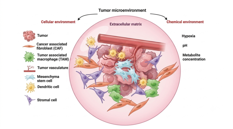
The tumor microenvironment components represents an intricate ecosystem surrounding cancer cells, composed of immune cells (such as macrophages, lymphocytes, and natural killer cells), stromal cells, blood vessels, and various signaling molecules. This environment plays a pivotal role in tumor development, metastasis, and response to treatments.
Introduction
The emergence of drug resistance in cancer is closely linked to mechanisms that reduce drug efficacy, with intratumoral heterogeneity and tumor microenvironment (TME) changes being major contributing factors. Heterogeneity of the tumor microenvironment components, a significant challenge to precision medicine, is traditionally attributed to genetic diversity, but recent findings reveal that epigenetic alterations from stochastic events and TME signals also play a crucial role.
In the context of preclinical oncology research, investigating and targeting thedifferents tumor microenvironment components is essential for the development of effective cancer therapies. Contract Research Organizations (CROs) specializing in oncology studies, particularly those developing murine cancer models, play a crucial role in advancing this research.
The Role of CROs in TME Research Using Murine Models
In the field of oncology, CROs provide essential expertise and infrastructure for conducting preclinical studies on the TME. The use of murine models allows researchers to closely replicate the human cancer environment, providing valuable insights into how the tumor microenvironment components can influence tumor behavior and treatment outcomes.
By leveraging murine models, CROs help test novel therapeutic strategies aimed at the TME before they move to clinical trials. These models are particularly important for studying immune interactions, stromal cell dynamics, and the role of various tumor microenvironment components such as tumor associated macrophages (TAMs), cancer-associated fibroblasts (CAFs), and the intratumoral microbiota.
Key Tumor Microenvironment Components in Preclinical Research
1) Tumor-Associated Macrophages (TAMs)
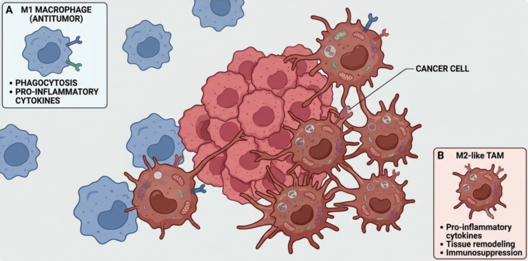
TAMs are critical players i1) n the TME, influencing the immune response and tumor progression. They can adopt different phenotypes, either promoting or inhibiting tumor growth. As the tumor progresses, TAMs often shift to a protumor M2 phenotype, supporting angiogenesis and metastasis while suppressing anti-tumor immune responses.
M2 TAMs inhibit cytotoxic T cell activity, promote regulatory T cell expansion, and support tumor proliferation, angiogenesis, and metastasis. Conversely, M1 TAMs are involved in phagocytosing cancer cells and mediating tumor destruction. Increased TAM abundance is often linked to poor patient outcomes and resistance to checkpoint inhibitor therapies, positioning TAMs as crucial prognostic biomarkers and therapeutic targets.
Preclinical models, particularly in murine systems, are invaluable for studying how TAMs can be targeted to enhance the efficacy of treatments like immune checkpoint inhibitors.
2) Cancer-Associated Fibroblasts (CAFs)
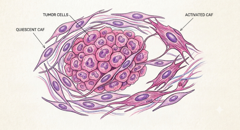
CAFs are another major component of the tumor stroma. They play a role in remodeling the extracellular matrix and regulating immune cell infiltration. CAFs are often associated with tumor progression and resistance to therapies, making them a significant focus in TME-targeted research.
Recent advancements in CAF-targeted therapies aim to deplete CAFs, mitigate their immunosuppressive functions, or reprogram them to a less active state. However, challenges remain in identifying specific CAF markers, understanding CAF subpopulations during tumor progression, and developing agents that target CAFs without affecting normal stromal cells.
CROs that specialize in preclinical oncology studies are at the forefront of testing therapies aimed at modulating CAF activity to inhibit their tumor-promoting functions while preserving normal tissue integrity.
3) Intratumoral Microbiota
The presence of microorganisms within tumors, collectively known as the intratumoral microbiota, has emerged as a novel area of research. These microorganisms can influence local immune responses and affect tumor growth and therapeutic outcomes.
Preclinical models, including those used by CROs, are instrumental in exploring how the microbiota contributes to tumor biology and in testing therapies that modulate microbial communities to improve cancer treatment outcomes.
Advancements in Preclinical Models for TME Research
Recent advancements in preclinical oncology research have allowed for more sophisticated studies of the tumor microenvironment components, particularly through the use of murine models. These models provide a controlled environment for studying the interactions between cancer cells and the various components of the TME. The precision offered by these models is critical for testing experimental therapies aimed at altering the TME to improve patient outcomes.
Many CROs working in the oncology space are integrating new technologies such as advanced imaging and flow cytometry to monitor tumor growth and immune cell activity in real time. These innovations enable a deeper understanding of how therapies interact with the TME and help accelerate the development of more effective treatments.
Conclusion: The Critical Role of the TME in Preclinical Oncology Research
The tumor microenvironment components are a complex and evolving area of study that has significant implications for understanding cancer progression, resistance and developing novel therapies. CROs specializing in preclinical oncology research, particularly those employing murine models, play a crucial role in advancing our understanding of the TME. These models provide valuable insights into the interactions between tumor cells, the immune system, and the surrounding stroma, allowing researchers to test and refine therapeutic strategies before they enter clinical trials.
As the field of TME research continues to evolve, the development of more predictive preclinical models will be essential in driving the next generation of cancer treatments.
Contact our Team to learn more
FAQ
Extracellular matrix (ECM) proteins, including collagens, laminins, and fibronectin, form a structural network that surrounds the malignant cells. Physical support is offered to tissue, and chemical signaling pathways are influenced by these structural molecules. Cancer progression is frequently supported when matrix stiffness is increased by altered collagen deposition. Biochemical signals are transmitted through specific receptors, which helps control cellular growth and movement. Tissue reorganization is facilitated when the ECM is continuously broken down by enzymes. Specialized testing platforms are utilized to evaluate how treatments affect these structural interactions. Therapies aimed at matrix remodeling are evaluated using these analytical tools.
New vascular pathways are generated through angiogenesis, which is stimulated by signaling molecules such as VEGF. Oxygen supply and nutrient transport are provided to growing masses via these newly formed vessels. Poor structural integrity is frequently displayed by tumor vasculature, which leads to leaky walls and irregular blood flow within the tissue. High fluid pressure is created in the surrounding tissue, which limits drug penetration. Vessel normalization strategies are studied to improve drug delivery efficiency. Immune cell entry into malignant regions is also altered by these vessel changes. Vascular patterns are analyzed in preclinical models to assess anti-angiogenic therapies.
Distinct immune populations, including T lymphocytes, natural killer cells, macrophages, and myeloid-derived suppressor cells, are found in the surrounding tissue. Antitumor responses are carried out by cytotoxic T cells, but their activity is often restricted by immunosuppressive signals. Tumor-associated macrophages (TAMs) are frequently altered to adopt an M2 phenotype, which supports tumor growth and suppresses immunity. Regulatory T cells and suppressor cells are recruited to prevent effective immune attacks. Immune checkpoint molecules, such as PD-1 and CTLA-4, are expressed to inhibit cell-mediated Destruction. Immunotherapy effectiveness is tested by profiling these cellular subsets. Drug candidates are evaluated using specialized cellular profiling assays.
Cancer-associated fibroblasts (CAFs) are activated by chemical signals within the local tissue. Growth factors, cytokines, and extracellular matrix components are actively secreted by these specialized cells. Physical barriers to drug entry are formed when CAFs deposit dense collagen fibers. Immunosuppressive signals are released by CAFs, which hinders active immune responses. Tumor invasion and metastasis are promoted through the continuous release of matrix-degrading enzymes. Cross-talk between CAFs and cancer cells is studied to find potential therapeutic targets. Inhibitors targeting CAF activation pathways are tested in preclinical oncology assays.
Hypoxic conditions are established when rapid cell growth outpaces the local blood supply. Hypoxia-inducible factors (HIFs) are activated in response to low oxygen levels, which triggers angiogenesis and altered metabolism. Increased lactic acid production leads to local tissue acidification. Immune cell function is impaired in acidic, low-oxygen areas, which helps cells evade destruction. Metastatic potential and resistance to radiation therapy are enhanced under hypoxic stress. Oxygen and pH variations are monitored in experimental models to evaluate treatment responses. Metabolic targeting drugs are tested using controlled oxygen testing conditions.
Cytokines, chemokines, and growth factors are released by stromal and malignant cells to coordinate local activities. TGF-beta, IL-6, and CXCL12 are secreted to suppress immune activity and promote cell survival. Paracrine signaling loops are formed between different cell types, which drives continuous tissue remodeling. Chemokine gradients are established to attract specific suppressor cells into the surrounding tissue. Signal transduction pathways are triggered upon binding to target cell surface receptors. Intercellular communication is measured using multiplex protein analysis tools. Interruption of these signaling networks is evaluated as a therapeutic strategy.
Physical barriers, such as dense ECM and high interstitial pressure, are created to reduce drug absorption. Immunosuppressive populations, including TAMs and suppressor cells, are recruited to protect malignant cells from immune-mediated destruction. Hypoxic regions are formed, which reduces the effectiveness of therapies that require oxygen to generate cytotoxic effects. Survival pathways are activated in cancer cells by signals received from surrounding stromal cells. Dormancy is induced in subset cells, which allows them to survive targeted chemical treatments. Complex multi-cellular models are used to test strategies for overcoming these resistance mechanisms.
Advanced laboratory systems, such as co-culture models, organoids, and humanized mouse models, are constructed to imitate local tissue interactions. Human immune populations are engrafted into immunocompromised mice to study human-specific responses. Spatial organization and cell-to-cell communication are recreated using 3D microfluidic devices. Drug distribution and cellular recruitment are measured within these specialized testing platforms. Model selection is guided by the specific therapeutic mechanism being evaluated. Tumor-stroma interactions are monitored over time to gather detailed activity data. Analytical assays are applied to evaluate drug efficacy in realistic tissue settings.
Target engagement and therapeutic efficacy are altered by complex stromal interactions surrounding malignant cells. Standard cell cultures fail to reflect the physical barriers and immunosuppressive conditions present in living tissue. Drug penetration, immune activation, and matrix remodeling are assessed using complex tissue models. Predictive data regarding combination therapies are provided by evaluating stromal responses. Safety and efficacy profiles are clarified before clinical testing begins. Biomarkers associated with response or resistance are identified through thorough tissue profiling. Preclinical testing protocols are tailored to include these essential microenvironmental measurements.
