

INTRODUCTION
The use of tumor cells as in vivo tumor models is a commonly used approach in biomedical research to study tumor development, progression, and response to
treatment. Tumor models recreate a three-dimensional environment closer to reality than cells growing in 2D in vitro. They enable to better reproduce the conditions present in patients’ tumors [1][2] and to better understand the mechanisms of tumor formation and progression. These models are based on tumor in vitro cell culture to be grown in large quantities [1] and injected into animal models such as immunocompromised mice [3]. Tumor models play a crucial role in the development of new treatments, enabling their efficacy, pharmacokinetic parameters, and potential side effects to be studied before they are tested in clinical trials [4]. As such, they are essential tools in the development of new anti-cancer treatments, such as Antibody Drug Conjugates (ADCs), bispecifics, Imune Checkpoint Inhibitors (ICI),…. To create an in vivo tumor model, many tumor cells are needed. The number of cells required for each experiment can run into the hundreds of millions.
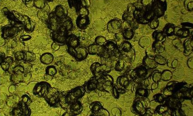
A collaborative effort between Antineo and Carroucell has harnessed the potential of Carroucell cutting-edge technology for the large-scale amplification of diverse tumor cell lines. The overarching goal is to address and alleviate the spatial constraints and economic burdens associated with the preparation of animal tumor models, particularly for Antineo
METHODS
In vitro cell culture:
SKOV3, Hs746t and MDA-MB-175 cells were transferred from Antineo to Carroucell. SKOV3 cells were cultured in RPMI medium (Thermo Fisher Scientific, Illkirch, France) supplemented with 10% fetal calf serum. Hs746t cells were cultured in DMEM medium (Thermo Fisher Scientific, Illkirch, France) supplemented with 10% fetal calf serum. MDA-MB-175 were cultured in DMEM-F12 medium (Thermo Fisher Scientific, Illkirch, France) supplemented with 20% fetal calf serum. All media were supplemented with 100 units/mL penicillin and 100 μg/mL streptomycin, respectively.
Microcarriers
Carroucell FlexiGrowTM disc microcarriers (batch 01-09-2023) with respective diameters of 85 and 275 μm were used. Those microcarriers were synthetized with Carroucell patented Sol-Gel process. At the end of the synthesis, microcarriers were functionalized by the inclusion of positive charges at their surface. Microcarriers sterility was obtained by autoclaving for 30 minutes at 121°C, 1.2 PS
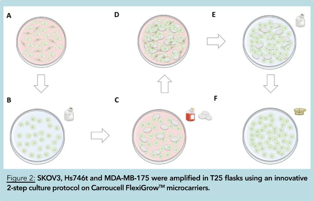
(A): 3.6 x 10⁵ SKOV3, 3 x 10⁵ Hs746t and 1 x 10⁶ MDA-MB-175 cells were seeded in individual T25 culture treated flasks, respectively. Cells were incubated for 4, 8 and 3 days respectively at 37°C, 80% humidity, and 5% CO2. The final volume of medium in each flask was 5 mL.
(B): At the end of the culture, cells were washed with PBS and detached by a 5-minute incubation with 1 mL Trypsin —EDTA. Cell quantity and viability were quantified after Trypan Blue staining using automated cell counting (LUNA II – Logos Biosystems, South Korea).
(C) and (D): Following quantification, total SKOV3, Hs746t and MDA-MB-175 cell amount was seeded again in the same T25 flflask supplemented with Carroucell FlexiGrowTM microcarriers. The ratio cells/microcarriers was set to 15 mg per 106 cells. CarrouceFlexiGrowTM microcarriers had diameters of 85 and 265 μm, respectively. Both SKOV3 and MDA-MB-175 cells were cultured for 4 additional days on Carroucell FlexiGrowTM microcarriers. Hs746t were cultured for 8 additional days on Carroucell FlexiGrowTM microcarriers. Final volume of medium in each flask was adjusted to 10 mL.
(E): At the end of the 2-step culture protocol on microcarriers, cells were washed in PBS and harvested by a 10-minute incubation with 2 mL Trypsin — EDTA.
(F): 4 mL of medium was added, and cells were separated from the microcarriers by filtration. 40 μm and 100 μm Corning© Cell Strainers were used for microcarriers with 85 and 265 μm diameters, respectively. Total cell quantity and viability were quantified after Trypan Blue
staining and compared to the ones obtained without microcarriers.
RESULTS
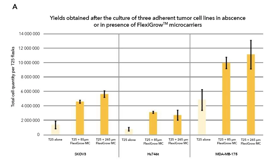
By applying Carroucell innovative 2-step protocol for adherent cell amplification in static culture environment, 4.6 x 10⁶ and 5.6 x 10⁶ SKOV3 cells were obtained per T25 on 85 μm and 265 μm FlexiGrowTM microcarriers, respectively. This is 3.3 and 4.1 times more than in classic culture protocol in T25 alone.
In comparison 7.6 x 10⁵ and 2.6 x 10⁶ Hs746t cells were obtained in absence (T25 alone) and in presence of 265 μm FlexiGrowTM microcarriers. In presence of 85 μm FlexiGrowTM microcarriers, 3.1 x 10⁶ Hs746t cells were obtained. This is 4.1 times more than in T25 alone.
Finally, 4.8 x 10⁶, 9.9 x 10⁶ and 11.1 x 10⁶ MDA-MB-175 were obtained in absence, or in presence of 85 μm and 265 μm FlexiGrowTM microcarriers, respectively. In this last condition, 2.3 times more cells were obtained in comparison with the T25 alone.
In this study, using an innovative 2-step culture protocol, an in vitro cell culture of adherent tumor cells were seeded on FlexiGrowTM microcarriers after a preamplification in the culture flask alone. For each million cells that were seeded, flasks were supplemented with 15 mg of FlexiGrowTM microcarriers. This optimal cell/microcarriers ratio has been optimized by Carroucell and showed excellent results for numerous types of adherent cells. This ratio permits to significantly increase the adherence surface available for the cells while preserving an optimal culture homogeneity.
Noteworthy, the culture on FlexiGrowTM microcarriers resulted in different outcomes according to the cell types and the microcarriers diameters. SKOV3, Hs746t and MDA-MB-175 cells are three different tumor cell types, isolated from three different organs, and characterized by completely different phenotypes and functionalities. Adapting FlexiGrowTM microcarriers size is therefore a crucial strategy to:
– Optimize cell interaction with microcarriers.
– Optimize the formation of cell-microcarriers tridimensional structures,
– Favor the optimization of the total adherence surface provided by both the culture flask and the microcarriers.
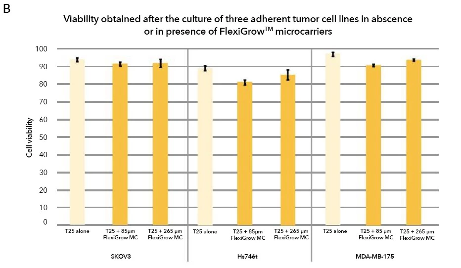
Furthermore, post-amplifification on Carroucell FlexiGrowTM microcarriers, the viability of cells exceeded 80%. This noteworthy outcome attests to the compatibility and supportive nature of the microcarriers culture protocol, ensuring the preservation of cell health and functionality throughout the amplification process.
The adherence to stringent viability thresholds reaffifirms the reliability and reproducibility of the Carroucell protocol. This emphasizes its potential for advancing cell culture methodologies in Oncology. More specifically for Antineo, such approaches aim to provide turn-key protocols while reducing resources dedicated to cell amplification processes and conserving the high robustness of the proposed in vivo
cancer models.
CONCLUSION
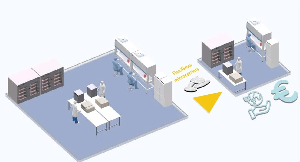
The utilization of Carroucell’s amplification process on FlexiGrowTM microcarriers initiates a transformative phase marked by significant efficiency gains. This innovative strategy ensures a 4-fold reduction in spatial requirements, a 2.5-fold decrease in consumables (encompassing media, trypsin, and plastic waste), and a 2-fold decrease in the time required by the technical team, as illustrated in Figure 4. This remarkable optimization of resources dedicated to cell culture is seamlessly achieved without disrupting established production chain practices.
Essentially, these strategic measures empower Antineo to reallocate a substantial portion of resources toward their core business: delivering resilient and versatile animal models in oncology. This streamlined operational approach not only minimizes costs but also provides Antineo with the agility and resources required to strengthen their position as pioneers in delivering cutting-edge solutions for oncology research. Carroucell’s approach revolutionizes resource utilization in cell culture, ensuring unparalleled efficiency, cost-effectiveness, and steadfast support for the core objectives of esteemed partners such as Antineo.
REFERENCES
[1] A. M. Nagornykh, M. A. Tyumentseva, A. I. Tyumentsev, et V. G. Akimkin, « Protocol for chronic hepatitis B virus infection mouse model development by patient-derived orthotopic xenografts », PLoS ONE, vol. 17, no 2, p. e0264266, févr. 2022, doi: 10.1371/journal.pone.0264266.
[2] V. Forest, L. Campos, M. Péoc'h, D. Guyotat, et J.-M. Vergnon, « [Development of an experimental model for the study of the effects of cryotherapy on lung tumours] », Pathol. Biol. (Paris), vol. 53, no 4, p. 199203, mai 2005, doi: 10.1016/j.patbio.2004.08.002.
[3] J. Jung, H. S. Seol, et S. Chang, « The Generation and Application of Patient-Derived Xenograft Model for Cancer Research », Cancer Res. Treat. Off. J. Korean Cancer Assoc., vol. 50, no 1, p. 110, janv. 2018, doi: 10.4143/crt.2017.307.
[4] A. S. T. Smith, J. Macadangdang, W. Leung, M. A. Laflamme, et D.-H. Kim, « Human iPSC-derived cardiomyocytes and tissue engineering strategies for disease modeling and drug screening », Biotechnol. Adv., vol. 35, no 1, p. 7794, janv. 2017, doi: 10.1016/j.biotechadv.2016.12.002.
[5] le Clainche T, Moisan A, Coll JL, Martel-Frachet V (2021) The disc-shaped microcarriers: a new tool for increasing harvesting of adipose-derived mesenchymal stromal cells. Biochem Eng J 174:108802. doi: 10.1016/j.bej.2021.108082
Download the application note here: Application note AntineoCarroucell 2024
Contact our Team to learn more
FAQ
Over 150 tumour models originating from human and murine sources are made available to evaluate candidate compounds. Research projects are supported across both solid and hematological cancers. In vitro cell culture assays are tailored to individual study requirements to streamline lead selection. Preclinical animal studies are thereby optimized in a cost-effective manner. Experimental designs can be aligned with specific therapeutic objectives.
Dynamic cellular variations are tracked continuously in real time using dedicated Incucyte and XCellingence equipment. Cellular proliferation, structural alterations, and cell death rates are observed progressively. Drug toxicity, therapeutic efficacy, and cellular expansion are recorded during treatment. Continuous observation allows precise evaluation of biological changes over time.
30 original resistant tumour models have been generated to investigate therapy failure mechanisms. Resistant lines are established to test combination therapeutic strategies. These cellular tools support the study of acquired resistance against standard therapeutic agents. Novel strategies to counter treatment resistance are refined using these platforms.
End-point assays are carried out at defined intervals to assess compound efficacy. Apoptosis, tumour growth inhibition, and immune reactions are measured systematically. Key biomarkers are analyzed to determine therapeutic actions across treatment stages. These controlled evaluations assist in confirming compound performance before animal trials.
Multidimensional cellular profiling is conducted using flow cytometry platforms equipped with up to 5 lasers. Biomarkers are quantified via 29-marker panels to characterize cell populations. Off-the-shelf or customized panels are deployed based on study goals. High-dimensional profiling allows precise evaluation of cellular responses.
In vitro cell culture systems are employed to assess novel immunotherapeutic modalities. Monoclonal antibodies, bispecific molecules, immuno boosters, and CAR-T cell therapies are tested. Immuno-oncology interactions and immune responses are measured in controlled environments. Microenvironment dynamics are thoroughly examined prior to in vivo testing.
Three-dimensional models are constructed by combining cancer cells with fibroblasts or immune cells. Tissue architecture and cell-cell interactions are reproduced accurately. Realistic models improve the evaluation of drug toxicity and efficacy. Screening predictability is enhanced while animal usage is minimized.
Patient-derived tumour cells are integrated into bioprinted structures to mirror clinical conditions. Individualized tumour profiles are recreated for targeted therapeutic testing. Treatment responses are assessed under conditions close to clinical reality. Screening efficiency is improved for personalized therapy evaluation.
In vitro cell culture assays accelerate candidate selection prior to animal studies. Efficacy and toxicity profiles are established quickly through medium-throughput screening. Studies are adapted to meet precise research requirements efficiently. The transition from early discovery to clinical development is facilitated smoothly.
