

Assessing Tumoral Volume in a Murine Colorectal Cancer Model Using MRI Technology
INTRODUCTION
Preclinical models, such as murine models, are essential for evaluating the efficacy of therapeutic treatment in oncology research. Recently, novel murine models with surgically implanted orthotopic tumor allow a more accurate representation of human cancer than traditional subcutaneous models. Orthotopic models however require in vivo evaluation. This shift necessitates the use of non-invasive imaging techniques to monitor tumor growth, as caliper measurements used for subcutaneous tumors are no longer applicable.
Monitoring the tumoral volume or its follow-up is often imprecise. It is also difficult to attribute weight changes to tumor progression or other confounding factors. One of the key endpoints in these studies is the assessment of tumoral volume, which allows researchers to monitor not only tumor growth but also response to treatments over time. MRI has emerged as a non-invasive and precise imaging technique providing detailed anatomical and functional information, making it particularly useful in oncology research.
This white paper, in collaboration with Hawkcell, meant as a proof of concept, discusses the pertinence of using MRI technology to assess tumoral volume in an orthotopic murine colorectal cancer model.
METHODS
Experimental Design
• Model: Female murine model (C57BL/6).
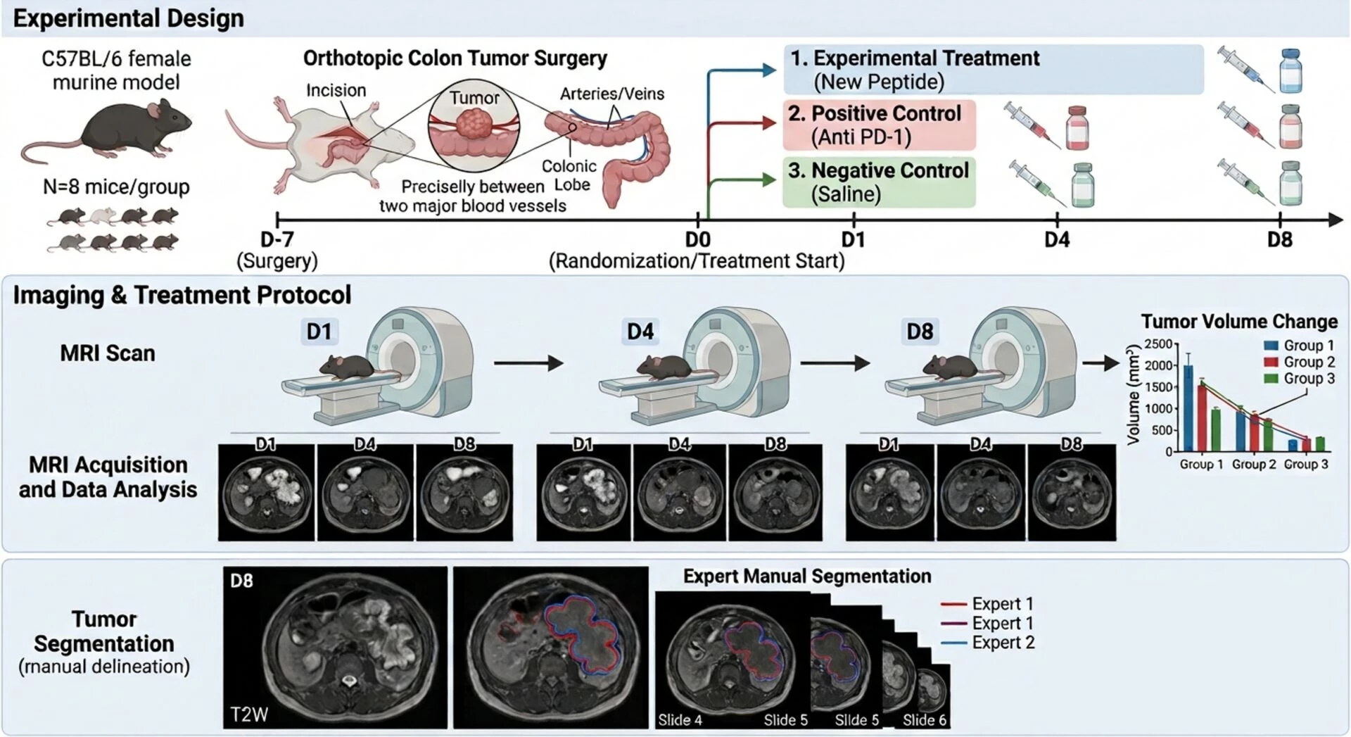
• Surgery: Colorectal tumor surgically implanted orthotopically on the colon segment between two blood vessels 7 days before D0.
Groups: Mice were divided into three groups:
1. Group 1 (Experimental Treatment): Treated with a new experimental peptide.
2. Group 2 (Positive Control): Treated with a known immune checkpoint inhibitor (Anti PD-1).
3. Group 3 (Negative Control): Treated with saline solution (NaCl).
• Sample Size: 8 mice per group.
• Imaging: MRI data acquisition was performed at three time points (D1, D4, and D8) – to assess changes in tumoral volume.
MRI Acquisition and Segmentation
Each colorectal cancer model volume at D1, D4 and D8 were manually segmented, as automated algorithms do not perform well due to the irregular nature of tumor shape, by two experts, to ensure precise delineation of tumor limits.
RESULTS
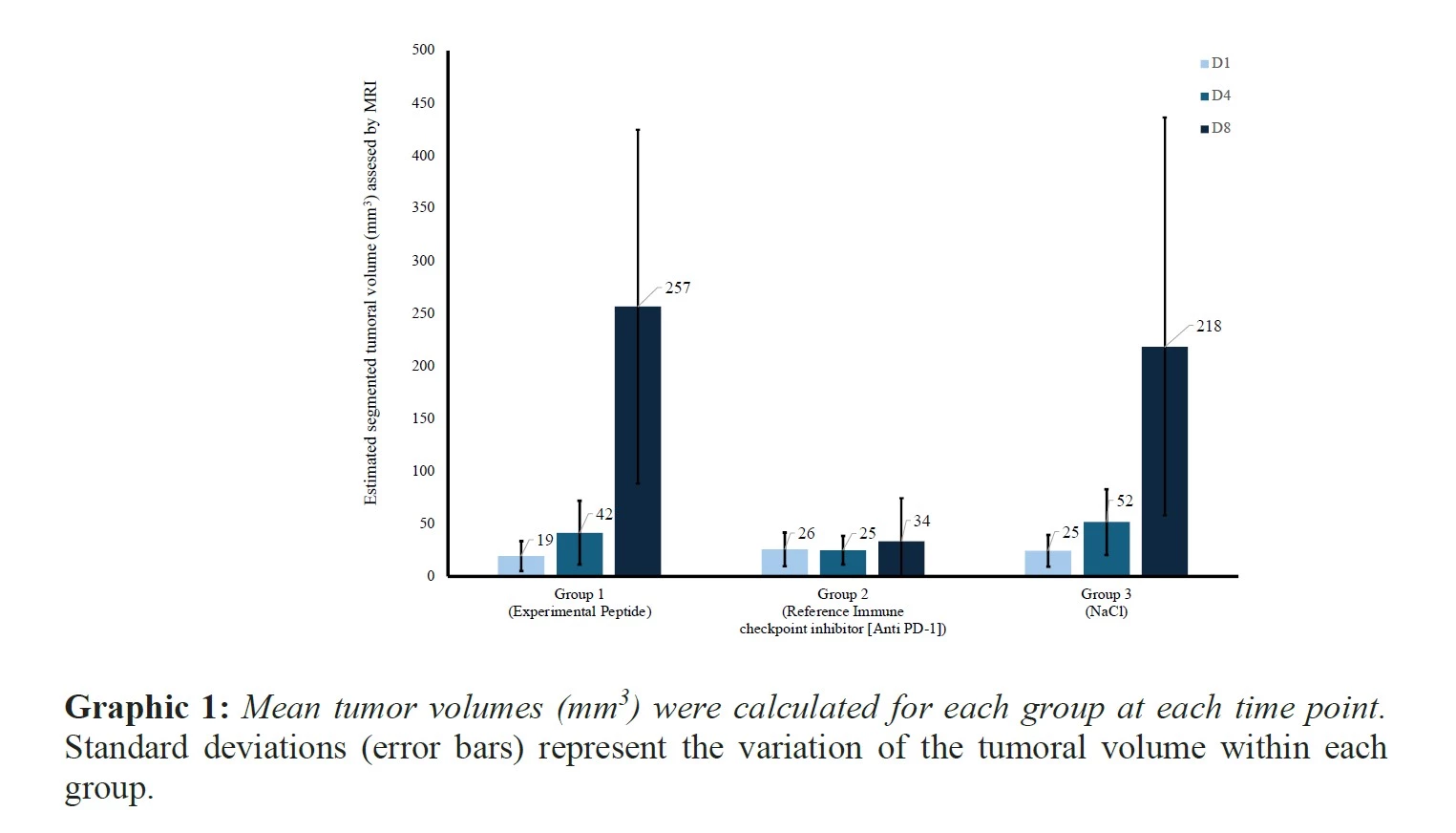
A significant increase in tumor volume was observed in Group 1 by Day 8, suggesting that the experimental treatment may be less effective in controlling tumor progression in rapidly growing tumor models, such as MC38 model (resistant or not), compared to the positive control group. Conversely, Group 2, treated with the well-established immune checkpoint inhibitor anti-PD-1, exhibited a slight reduction in tumor volume, demonstrating the efficacy of this control treatment in suppressing tumor growth. The NaCl control group displayed consistent tumor growth, with a substantial increase in volume by Day 8, confirming the aggressive nature of tumor progression in this untreated model.
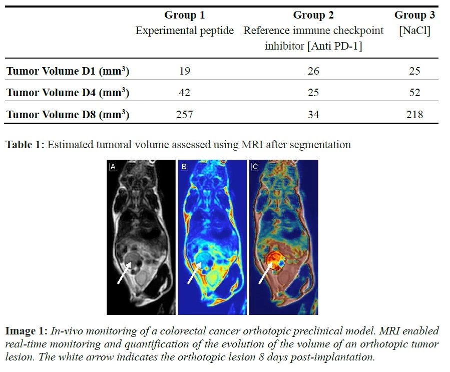
The use of MRI for measuring tumoral volume provides valuable data for early-phase drug development in cancer research. The ability to visualize and quantify tumor progression or regression offers critical insights into the efficacy of novel therapeutic agents. Moreover, the non-invasive nature of MRI means that researchers can obtain frequent, reliable measurements from the same animals, thereby increasing the statistical power of the study and reducing animal usage.
DISCUSSION & CONCLUSION
Our results underscore the invaluable role of MRI in tracking tumor growth over time without the need for invasive procedures. Compared to traditional methods like caliper measurements or histological analysis, MRI offers several key advantages:
• Precision: MRI’s 3D volumetric capabilities enable accurate measurement of irregularly shaped tumors, which are frequently observed in cancer models.
• Non-Invasive Nature: Unlike histological assessments that necessitate animal sacrifice at multiple time points, MRI allows for longitudinal studies in the contemporaneous cohort, minimizing variability and reducing the number of animals required.
• Early Detection: MRI’s sensitivity in detecting small tumor volumes as early as day 1 is crucial for evaluating the early efficacy of therapeutic interventions.
• These advantages make MRI an indispensable tool for preclinical research, providing valuable insights into tumor biology and the effectiveness of potential treatments.
The results from this colorectal cancer model (syngeneic) underscore the pertinence of using MRI technology for tumoral volume assessment. The high accuracy, non-invasive nature, and ability to monitor tumor growth longitudinally make MRI a gold standard for preclinical cancer studies. While the experimental treatment group exhibited significant variability in tumor response, the positive control group demonstrated the efficacy of an anti-tumoral agent, and the NaCl control group showed typical aggressive tumor progression. These findings reinforce the value of MRI in preclinical evaluations and its role in advancing the development of effective cancer therapies.
This white paper provides a foundation for further studies that can refine the experimental design, optimize the treatment regimen, and expand on the use of MRI in oncology research.
The MRI imaging is a very good complementary technique to luciferase bioluminescent imaging of live animal.
Contact our Team to learn more
Download the new white paper here.
FAQ
Tumour progression within an orthotopic colorectal cancer model is monitored non-invasively with high-field magnetic resonance imaging. Images are collected regularly. Internal tissue changes are tracked across multiple time points. Soft tissues are differentiated clearly by this non-ionising imaging technique without surgical intervention. Detailed anatomical structures inside the abdominal cavity are exposed clearly. Primary lesions and secondary growths can be quantified with high precision. Longitudinal studies are supported because the same animal is scanned repeatedly over several weeks. Drug responses to novel agents are assessed by comparing volume changes across treatment cohorts. Precise diagnostic information is obtained while animal welfare is preserved. Image data are subsequently processed to calculate exact total lesion volumes.
Orthotopic implantation into the caecum wall is chosen because natural organ microenvironments are recreated accurately. Subcutaneous models lack this anatomical relevance. Tumour growth, vascularisation, and local invasion are mimicked more faithfully in the primary organ site. Metastatic spreading to distant organs like the liver can also be monitored effectively. Internal development is observed non-invasively through abdominal magnetic resonance imaging scans. Interactions between cancer cells and surrounding stromal tissues are preserved in orthotopic settings. Experimental compounds can thus be tested in conditions that reflect natural pathology closely. Better predictive value for clinical trials is attained through this physiological context. Research outcomes are thereby improved through realistic tissue interactions.
Deep internal organs inside small animals cannot be reached by mechanical calipers. Magnetic resonance imaging is relied upon for accurate non-invasive measurement of internal tumours. Precise three-dimensional volume measurements are generated instead of simple superficial estimations. Asymmetrical growths and deeply seated abdominal masses are detected accurately. Early stage lesions are identified well before palpable nodules appear on the surface. Radiation exposure is completely avoided during magnetic resonance procedures. Repeated imaging sessions are conducted safely without harm to subject tissues. Accurate anatomical resolution is provided for both primary tumours and metastatic deposits. Quantitative data are extracted reliably to track therapeutic efficacy over time.
Liver and lymph node metastases originating from an orthotopic colorectal cancer model are identified using high-resolution magnetic resonance sequences. Contrast agents are sometimes administered intravenously to boost image clarity. Microscopic metastatic foci are detected within abdominal organs at early developmental stages. Tissue changes in the liver parenchyma are monitored sequentially without sacrificing host animals. Quantitative volumetric analysis is performed on all identified secondary lesions. Disease dissemination patterns are mapped comprehensively across the entire abdominal cavity. Treatment efficacy against metastatic spread is evaluated by comparing scan results between control and treated groups. Reliable tracking of advanced systemic disease is maintained throughout testing.
Raw magnetic resonance data are converted into detailed digital images for computational analysis. Target organ boundaries and tumour margins are delineated using specialized image segmentation software. Total lesion volumes are calculated automatically by summing contiguous slice areas. Signal intensity variations are analysed to evaluate internal necrosis or fluid accumulation within malignant tissues. Differences in soft tissue contrast are measured quantitatively across experimental groups. Standardised digital protocols are followed to ensure consistent analysis between different study cohorts. Final visualisations are compiled into comparative graphs and three-dimensional structural renders. Data integrity is guarded through rigorous automated calibration steps.
Therapeutic responses within a colorectal cancer model are determined by measuring alterations in tumour volume over time. Regression or stabilization of tissue growth is observed directly in serial scans. Internal changes such as necrosis or altered vascular permeability are detected before structural shrinkage occurs. Drug candidate efficacy is compared against control vehicles using standardized image metrics. Scan data are integrated with biomarker assessments to verify biological mechanism. Non-responders are identified quickly during early treatment cycles. Precise volumetric endpoints are obtained without requiring terminal tissue collection at intermediate stages. Study design efficiency is enhanced while animal usage numbers are minimized.
Standard anatomical evaluation of abdominal structures is frequently performed without exogenous contrast media. Endogenous tissue contrast between healthy muscle, fat, and tumour mass is sufficient in high-field magnetic resonance systems. Gadolinium-based contrast agents are administered when vascular perfusion or membrane permeability must be measured specifically. T1-weighted and T2-weighted scan sequences are adjusted to highlight distinct pathological features. Tumour borders are defined clearly against surrounding normal caecal tissue using optimized acquisition parameters. Unnecessary chemical exposure is avoided when native tissue contrast yields adequate diagnostic detail. Protocol selection is tailored according to specific analytical requirements.
Animal welfare during magnetic resonance procedures is maintained through continuous physiological monitoring. Inhalation anaesthesia is supplied during the entire scanning period to prevent movement artifacts. Body temperature is controlled using heated water pads or warm air circulation systems. Respiratory rates are tracked continuously with pneumatic sensors placed on the abdomen. Scan durations are minimized through optimized fast-acquisition imaging sequences. Rapid recovery from anaesthesia is routinely observed post-scan. Subject stress is minimized by reducing handling frequency and maintaining comfortable housing conditions. Ethical compliance is guaranteed through strict adherence to approved animal care protocols.
Syngeneic models are established using immunocompetent mouse cell lines such as CT26 or MC38. Murine cancer cells are injected directly into the caecum wall of matching host strains. Intact immune systems allow the study of immunotherapies and check-point inhibitors. Tumour microenvironment interactions involving native immune cells are preserved fully. Growth rates and metastatic capabilities are monitored using routine magnetic resonance imaging protocols. Responses to targeted biological therapies are assessed in fully immunocompetent hosts. Preclinical findings are translated more effectively to human clinical scenarios. High reproducibility in tumour engraftment is achieved across test cohorts.
