

ANTIBODY DRUG CONJUGATES (ADCS) : TRANSFORMING TARGETED CANCER THERAPIES
Abstract
Antibody drug conjugates (ADCs) represent a major advance in the fight against cancer. These innovative therapies act as veritable “homing missiles”, directly targeting cancer cells while sparing healthy tissue. By combining the specificity of monoclonal antibodies with the potency of cytotoxic agents, ADCs are emerging as one of the most promising forms of targeted therapy, offering patients more effective and less toxic treatment options.At Antineo, we are committed to supporting our customers in demonstrating the efficacy and safety of their ADCs, in order to make cancer treatments more precise and patient-friendly
What are Antibody Drug Conjugates ?
ADCs are an innovative class of biomedicines composed of three essential elements :
- Monoclonal antibody (mAb) : designed to specifically target antigens overexpressed on the surface of cancer cells
- Cytotoxic agent (payload) : a powerful chemotherapeutic agent attached to the antibody, designed to destroy cancer cells
- Chemical linker : this ensures the connection between the antibody and the drug, guaranteeing the stability of the complex in the bloodstream and enabling release of the cytotoxic agent only inside the cancer cell
This approach enables chemotherapy to be administered directly to the tumor, limiting systemic exposure and reducing the side effects usually associated with conventional treatments.
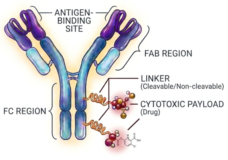
Antibody Drug Conjugates rely on the use of powerful cytotoxic agents to eliminate cancer cells. Below are listed the main classes of agents used as ADCs' payloads :
Auristatins (microtubule inhibitors) : Monomethylauristatin E (MMAE), Monomethylauristatin F (MMAF)
Maytansinoids (microtubule inhibitors) : DM1 (Maytansine derivative), DM4
Calicheamycin derivatives (DNA breakage-inducing agents) : N-acetyl-γ-calicheamycin
Camptothecins (topoisomerase I inhibitors) : SN-38 (active metabolite of irinotecan)
Pyrrolobenzodiazepines (PBDs) (DNA alkylating agents) : PBD dimers
Duocarmycins (DNA alkylating agents) : Duocarmycin derivatives
Indolino-benzodiazepines (DNA intercalating agents) : Tesirine derivatives
ADCs’ mechanism of action
ADCs operate in a targeted, multi-step process :
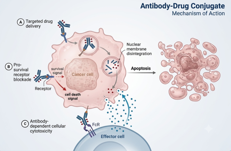
1.Recognition and binding : the ADC monoclonal antibody binds specifically to the tumor antigen present on the surface of cancer cells
2.Internalization : Once bound, the ADC is internalized by the cell via endocytosis.
3.Release of cytotoxic agent : After internalization, the linker is cleaved, releasing the cytotoxic agent directly into the cell.
4.Cellular destruction : The cytotoxic agent interferes with key processes in the cancer cell (DNA replication, cell division), leading to apoptosis (programmed cell death) and tumor shrinkage.
This targeted approach greatly reduces damage to normal, healthy cells, offering a more selective and effective treatment while minimizing side effects.
Major ADC approvals – clinical efficacy and safety
Recent clinical trials have led to the approval of several ADCs revolutionizing cancer treatment :
Brentuximab vedotin (Adcetris®) for lymphoma : Targeting the CD30 protein present on the surface of lymphoma cells, this ADC has demonstrated response rates in excess of 70% in patients with relapsed or refractory Hodgkin’s lymphoma
Trastuzumab emtansine (Kadcyla®) for HER2+ breast cancer : Combining trastuzumab with the cytotoxic agent emtansine, it significantly improved survival in patients who had failed previous treatment.
ADCs already approved show remarkable clinical efficacy, with high response rates and increased survival. The most common adverse effects remain manageable (fatigue, neutropenia, mild gastrointestinal disorders), while serious toxicities are rare thanks to the specificity of targeting.
Conclusion – Challenges and future prospects
Despite their potential, ADCs still face multiple challenges :
Tumor resistance : Some cancers develop resistance mechanisms, making ADCs less effective Residual toxicity : Even with a precise target, some ADCs may exhibit off-target toxicity.
Manufacturing difficulties : The production of ADCs is complex and costly, requiring advanced technologies.
However, the future of ADCs is promising, thanks to technological advances, the development of new therapeutic combinations (with immunotherapies or checkpoint inhibitors), and the extension of their application to solid cancers. ADCs represent a revolutionary advance in cancer treatment, enabling a more effective and better tolerated targeted approach.
With a deep understanding of ADCs' structure, mechanism of action, clinical successes, and the current challenges, we at Antineo are committed to help our customers advancing this transformative technology through preclinical phases.
Additional ressources
Dumontet et al. “Antibody drug conjugates come of age in oncology.” Nature reviews
Contact our Team to learn more
FAQ
Antibody drug conjugates are produced by joining a targeted monoclonal antibody to a toxic payload using a molecular linker. Tumour antigens are bound by the antibody portion. Meanwhile, destructive cellular processes are initiated by the payload once inside the target cell. Chemotherapy agents or radioisotopes are often chosen as payloads to destroy cancerous tissue. Structural stability is maintained by the linker until intracellular release is required. Target specificity and drug potency are united in these constructs. Biological effects are influenced by both the chosen antibody and the connected drug payload. Drug design must balance all three elements for optimal preclinical success.
Tumour destruction is achieved by antibody drug conjugates through targeted binding to specific surface proteins. Monoclonal antibodies bind to selective receptors expressed on tumour cell surfaces. The entire complex is taken into the cell via receptor-mediated endocytosis. After internalisation occurs, the chemical linker is cleaved inside lysosomal compartments. Free cytotoxic agents are then released directly into the cytoplasm. DNA damage or microtubule disruption is caused by the active toxin. Surrounding normal tissues are preserved because non-target cells lack these targeted surface proteins. High therapeutic indices are obtained through this localised activity.
In vitro and in vivo evaluations of antibody drug conjugates are performed in specialised preclinical laboratories. Target binding affinity is measured through binding assays. Cytotoxicity and internalisation rates are assessed in human cancer lines. Pharmacokinetic profiles are monitored in rodent models to determine drug stability. Efficacy studies are conducted using subcutaneous or orthotopic xenografts. Syngeneic mouse models are also utilized to evaluate immune interaction. Plasma clearance and off-target toxicity are measured to guide candidate selection. Detailed data sets are provided to support clinical trial applications.
Linkers for antibody drug conjugates are chosen based on required stability profiles in circulation. Cleavable linkers are designed to respond to intracellular enzymes or chemical conditions. Glutathione levels or acidic lysosomal environments trigger payload detachment. Non-cleavable linkers depend on total antibody degradation within lysosomes to release the active drug. Unintended toxicity is prevented by preventing premature cleavage in bloodstream plasma. Conjugate stability is tested across various biological fluids. Linker selection influences both target specificity and off-target side effects. Structural integrity must be maintained during systemic circulation.
Internalisation rate of antibody drug conjugates is measured using fluorescence microscopy and flow cytometry. Fluorescent dyes are attached to antibodies to track cellular uptake over time. Intracellular accumulation is quantified by comparing surface-bound signals with internalized signals. Quenching reagents are applied to remove non-internalized fluorescence on cell surfaces. Lysosomal co-localisation is verified using specific organelle markers. Quantitative metrics are produced to evaluate conjugate performance across target cell lines. Rapid uptake is preferred to ensure effective intracellular delivery of cytotoxic payloads.
Payload toxicity from antibody drug conjugates is evaluated using cell viability assays. Target-expressing cells and control non-expressing cells are exposed to varying drug concentrations. Cell death is quantified using luminescent or colorimetric metabolic indicators. Inhibitory concentration values IC50 are calculated to determine drug potency. Off-target cytotoxicity is checked to confirm target dependence. Bystander killing effects are also evaluated in co-culture systems. Free payload toxicity is compared directly against conjugated antibody efficacy. Accurate dose response curves are constructed to guide safe animal dosage.
Xenograft models are routinely used to assess antibody drug conjugates in vivo. Human cancer cells are implanted into immunodeficient mice to create target tumours. Patient-derived xenografts are also established to retain heterogeneous human tumour structures. Syngeneic rodent models are employed when immune system involvement is analyzed. Tumour growth inhibition is tracked by measuring tumour volume over several weeks. Animal body weight and health indicators are monitored to identify systemic side effects. Plasma samples are collected to analyze drug pharmacokinetics and clearance rates.
Bystander killing is observed when released payloads diffuse out of target cells into neighboring cells. Neutral cytotoxic molecules easily cross cell membranes after intracellular cleavage occurs. Adjacent tumour cells lacking surface target proteins are destroyed by these free drug molecules. Heterogeneous tumours are treated more effectively when bystander activity is present. However, potential systemic toxicity must be monitored if payload leakage occurs into circulation. Linker design influences whether bystander effects occur within solid tumours. Specific payload properties dictate membrane permeability after release.
Binding affinity of antibody drug conjugates is measured using surface plasmon resonance and cell-based assays. Binding kinetics are analyzed by exposing target antigens to various conjugate concentrations. Equilibrium dissociation constants Kd are determined to quantify binding strength. Flow cytometry is used to measure binding directly on live target cells. Binding properties of conjugated antibodies are compared with unmodified parent antibodies. Unaltered target binding confirms that chemical conjugation has not disrupted antibody structure. High specificity is required to avoid non-specific binding in non-target tissues.
