

Of the impact of the tumor microenvironment on acquired resistance: application note on colorectal cancer and anti-PD1 acquired resistance
Tumor microenvironment and cancer resistance
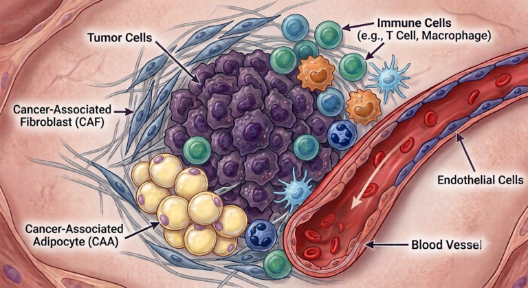
The tumor microenvironment (TME) is defined by the National Cancer Institute (NCI) as “the normal cells, molecules, and blood vessels that surround and feed a tumor cell. A tumor can change its microenvironment, and the microenvironment can affect how a tumor grows and spreads”[1]. Anderson et al., in 2020, also include the extracellular matrix component in the TME[2], and by citing Truffi et al.,[3] introduce the notion of a dynamic and reciprocal relationship between cancer cells and components of the TME to support cancer cell survival.
The TME has been proven to be linked to the occurrence of tumor resistance[4],[5] by playing a pivotal role in drug (small molecule or biologics) response by fostering immune evasion, altering drug availability, and facilitating cellular crosstalk that ultimately promotes resistance.[6]
Antineo's expertise in resistant models and the tumor microenvironment
Antineo's expertise in the development of secondary resistant oncology in vivo models has been used to investigate the impact of the tumor microenvironment on the occurrence and maintenance of the said resistance.
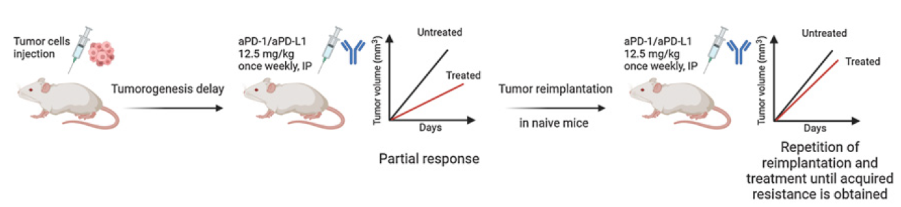
To this end, C56Bl6 mice have been subcutaneously injected with resistant colorectal cancer murine cells (MC38, part of Antineo's proprietary resistant model catalog), as described in the previous figure. The MC38 cells used are resistant to an immune checkpoint inhibitor, anti-PD1.
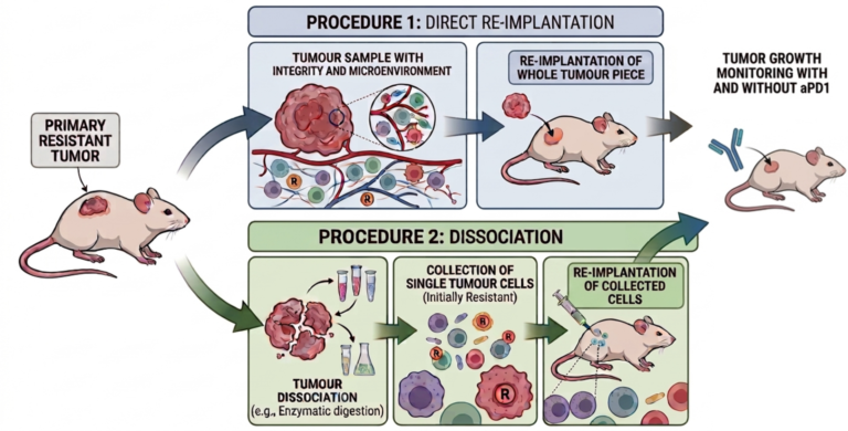
After collection of the formed tumor, 2 experimental procedures were performed.
- Direct re-implantation of the sampled tumor, with its whole integrity and tumor microenvironment
- Dissociation of the sampled tumor, collection of the tumor cells (initially resistant) and reimplantation in mice. This procedure destroys and discards the elements of the “natural” microenvironment.
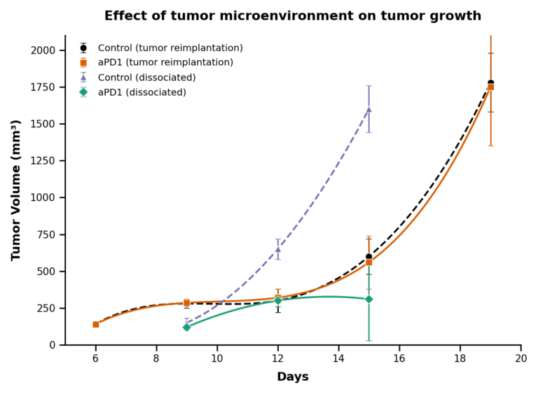
As the results show, the tumor cells reimplanted with the whole microenvironment (procedure 1) have kept the acquired resistance, as they are growing under the anti-PD1 treatment (orange full line) similar to the untreated group (black dashed lines).
After dissociation and loss of the tumor microenvironment, we observe a loss of the resistance of the cancer cells. Indeed, the tumor growth is almost completely inhibited by the anti-PD1 (green full line curve) compared to the dissociated control group (purple dashed line).
These results confirmed the critical role of the tumor microenvironment in the acquired resistance to specific drugs.
Such findings are also valid for in vitro models. When addressing resistance, the presence of the tumor microenvironment has to be recapitulated. In this case, tumoroids are the best option. To this extend, Antineo's 3D bioprinting tumoroids are specifically engineered to mimic the original tumor microenvironment as best as possible to provide an exploitable dataset to our customer.
Why does TME matter in drug development?
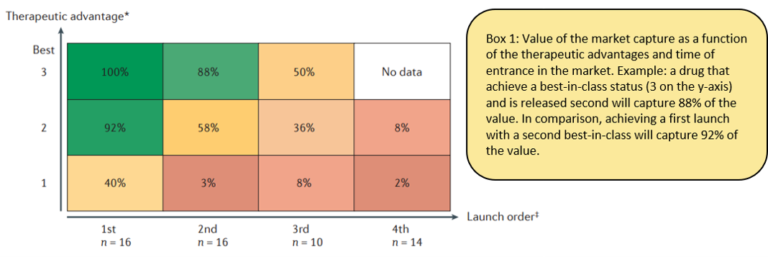
Schulze and Ringel made a meta-analysis on the market value taken in the case of a first-in-class or best-in-class[7]. In both cases, drug developers have a strong incentive, from the market itself, but also from the healthcare systems to bring added value with a new drug market launch.
This added value, in oncology, is either fewer side effects or in most cases, less relapse after remission of the primary tumor. This means developing a drug or combination that can act and treat resistant tumors is the key to market success.
Having access to not only cancer cells, but the recapitulation of the tumor in its whole complexity is key to accelerate and optimize the development of the most promising drug candidate, and future best/first in class
Contact our Team to learn more
FAQ
The tumor microenvironment is defined as the combination of normal cells, extracellular molecules, and blood vessels that surround and nourish a tumor cell. It is established that a tumor can alter its surrounding microenvironment. Conversely, the microenvironment itself can influence the growth and spread of the tumor. The extracellular matrix was later incorporated into this definition by Anderson et al.. A dynamic, reciprocal interaction between cancer cells and microenvironmental components is acknowledged to support cancer cell survival. Drug responses are affected because the microenvironment alters drug availability, promotes cellular crosstalk, and fosters immune evasion.
Colorectal cancer murine cells, designated as MC38, were selected for the preclinical evaluation. These cells belong to the catalog of proprietary resistant models developed by Antineo. The specific MC38 cells used in the experimental design are resistant to anti-PD1, an immune checkpoint inhibitor. To perform the study, the cell line was subcutaneously injected into C56Bl6 mice. Resistance models of this type are utilized to analyze how secondary resistance develops and is maintained within oncology studies. Through these models, broader mechanisms involving drug responses to small molecules and biologics can be studied effectively.
Tumor cells are initially injected into mice, after which a tumorigenesis delay is observed. Treatment is then administered via intraperitoneal injections of $\text{aPD-1}/\text{aPD-L1}$ at $12.5\text{ mg/kg}$ once weekly. A partial response is observed in the treated group compared with untreated controls. The resulting tumor tissue is harvested and re-implanted into naive mice. The weekly treatment regimen is repeated during subsequent re-implantations. This cycle of re-implantation and therapy is continued until acquired resistance is achieved. Tumor volume measurements are monitored continuously throughout the process.
When a primary resistant tumor piece is re-implanted directly, its whole structural integrity and microenvironment are preserved. Tumor volume growth is monitored over time under both treated and untreated conditions. The re-implanted tumor cells retain their acquired resistance to the treatment. Under anti-PD1 therapy, the tumor continues to grow at a rate similar to the untreated control group. By day 19, the tumor volume reaches approximately $1750\text{ mm}^3$ in both treated and control cohorts. Maintaining the intact microenvironment ensures that drug resistance is successfully preserved during tumor progression.
Dissociation of the primary resistant tumor is carried out through enzymatic digestion to isolate single cancer cells. This procedure destroys and discards the components of the natural tumor microenvironment. When these isolated cells are re-implanted into mice, a loss of drug resistance is observed. Tumor growth is almost completely inhibited by anti-PD1 treatment compared to the dissociated control group. By day 15, the average tumor volume of treated dissociated cells remains below 300mm3, whereas control tumors expand beyond 1500mm3. Thus, resistance is lost without the surrounding microenvironment.
The microenvironment must be recapitulated during in vitro testing to reflect accurate drug resistance mechanisms. Tumoroids are considered the most appropriate option for mimicking these complex cellular surroundings. Antineo's 3D bioprinting tumoroids are engineered to reproduce the original tumor microenvironment as accurately as possible. This structural recapitulation helps generate exploitable datasets for research applications. By maintaining realistic cellular surroundings, drug candidates can be evaluated more reliably outside of animal models. In vitro models lacking these structural components fail to preserve the acquired resistance observed in original tissues.
According to a meta-analysis by Schulze and Ringel, the timing of market entrance strongly dictates financial value capture. A first-in-class drug that achieves the highest therapeutic advantage score captures $100\%$ of the market value. If a product with the highest therapeutic advantage is launched second, its market value capture drops to $88\%$. A second-best drug launched first still captures $92\%$ of the value. Conversely, a third-choice drug launched second captures only $3\%$ of the value. Strong market incentives therefore exist for developers to launch effective therapies early.
Added therapeutic value in oncology is demonstrated either by reducing side effects or by preventing relapse after remission. Preventing tumor relapse requires therapies that can successfully target and eliminate resistant cancer cells. Market incentives and healthcare systems reward drugs that offer these verified clinical advantages. Developing single agents or combination therapies capable of treating resistant tumors is identified as a major requirement for commercial success. Access to the full complexity of a tumor, rather than isolated cancer cells alone, accelerates the identification of viable drug candidates. This comprehensive evaluation aids in creating first-in-class products.
The microenvironment encompasses diverse cell types surrounding the primary tumor mass. Endothelial cells line the adjacent blood vessels that supply the tumor tissue. Immune cells, including T cells and macrophages, are present within the matrix. Cancer-associated fibroblasts (CAF) surround the central cluster of cancer cells. Additionally, cancer-associated adipocytes (CAA) form a distinct cellular component of the surrounding matrix. Together with the extracellular matrix, these cells establish a dynamic structural network. This network interacts continuously with the tumor to support cellular survival and influence treatment response.
Preclinical studies and growth operations at Antineo are directed by designated leaders. Marie Tautou, PhD, serves as the Head of Preclinical Studies. Pierre Gaudriault, PharmD, PhD, serves as the Head of Growth Development. Inquiries regarding their preclinical models and proprietary tumoroid technologies can be directed to their central contact email address, contact@antineo.fr. Their team provides expertise on resistant oncology models and in vivo tumor microenvironment evaluations.
