Our panel of in vitro end-point studies assess the antitumoral effect of your lead compounds, as well as their mechanisms of action on cell cycle, or on the immune system. Our capacity to perform multiple colour flow cytometry helps you characterising your target cells, and indications.

Our offer includes

  • Determination of cytotoxicity and IC50
  • Synergy assays between compounds or with gold standards
  • Analysis of cell cycle mechanisms (apoptosis, stasis, proliferation, phases modifications)
  • Multiple colour flow cytometry
  • Immunology and immuno-oncology assays (T cells activation, CTL assay, etc)

In vitro cytotoxicity

In vitro cytotoxicity

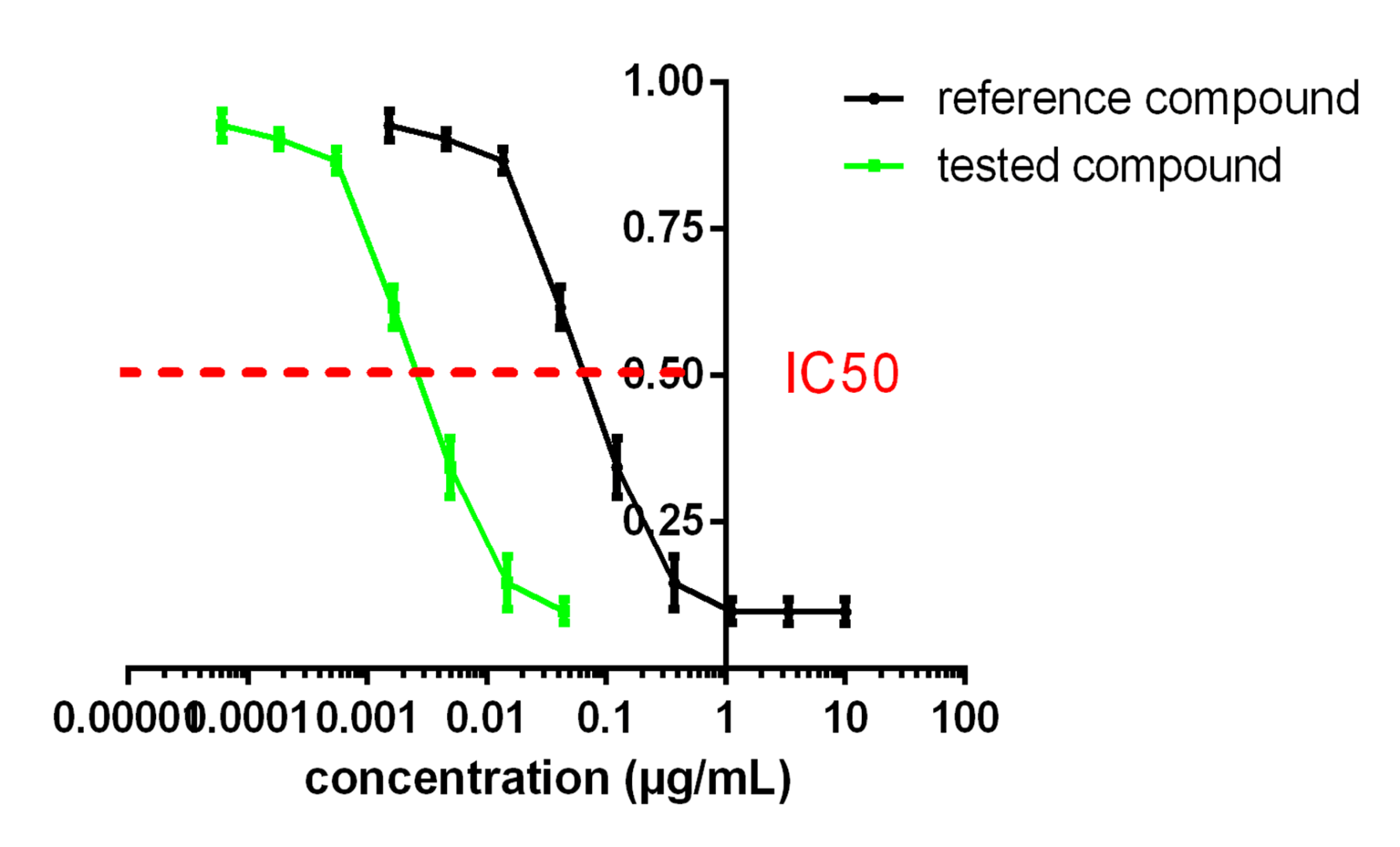
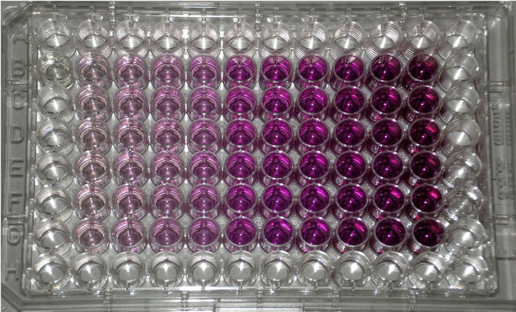
At Antineo, we perform MTT assays to assess the cytotoxic potential of your compounds. This efficient assay determines the IC50 of your compound on a given tumour model. The IC50 is the concentration for which the compound induces a 50% decrease in mitochondrial metabolism (correlated with cell survival) in comparison to control.

MTT assays are used to screen our bank of preclinical tumour models for sensitivity to your compound, but also to compare the ranges of sensitivity of a given model with the gold standard. They can also help you to screen for unwanted cytotoxicity.

The assay is performed with triplicate wells per concentration and the IC50 is calculated with the Compusyn software (ComboSyn Inc, USA).

Antineo is able to complete the data obtained in end-point MTT assays by continuous-monitoring assays (see continuous monitoring).ee continuous monitoring).

In vitro synergy assays

In vitro synergy assays

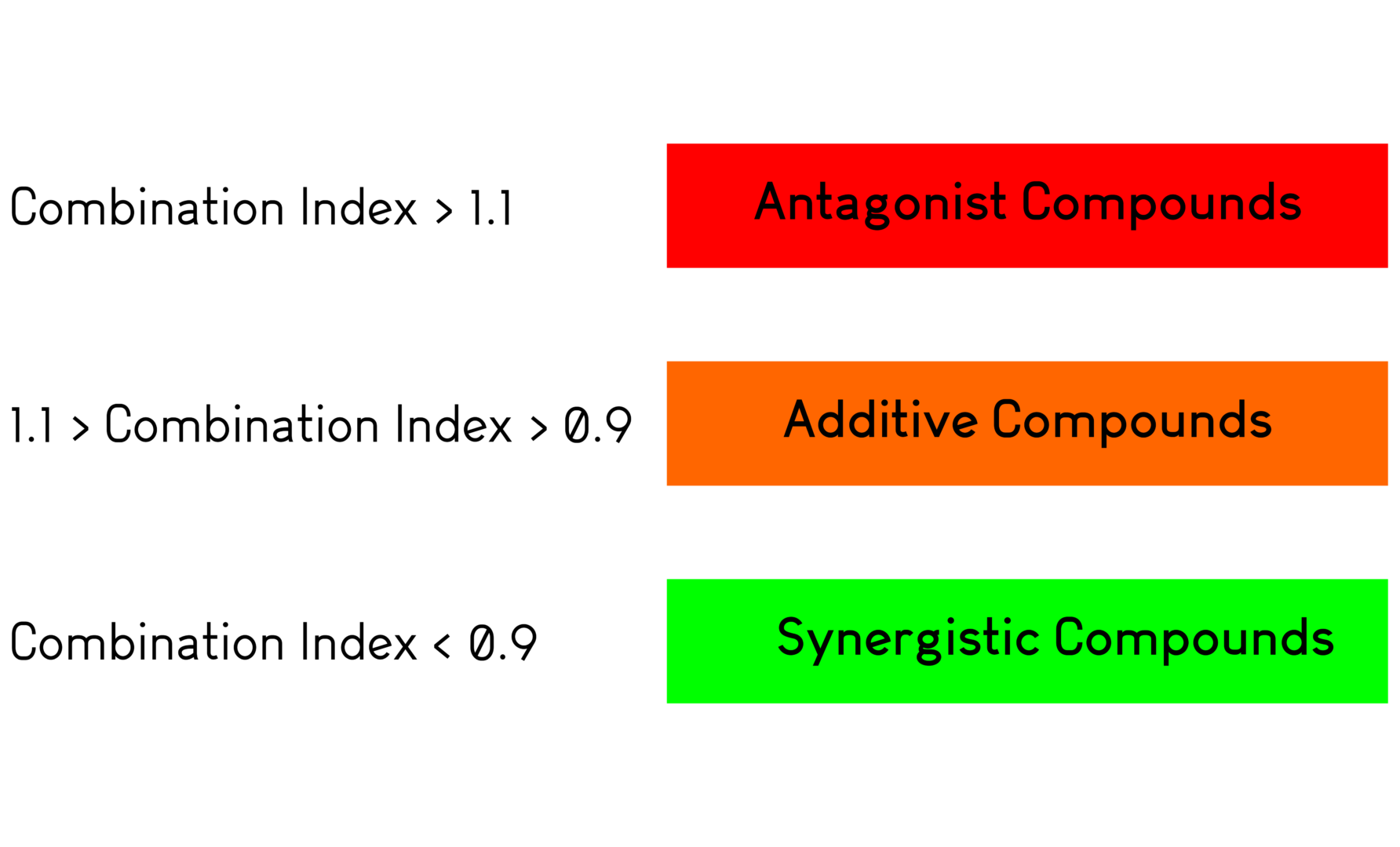
Antineo uses Compusyn software (ComboSyn, Inc., USA) to determine synergy or antagonism between anticancer agents, from IC50 data obtained by MTT assays. This software determines the IC50 of two compounds as well as the Combination Index (CI). The CI values indicate whether two agents are antagonistic, additive or synergistic in terms of cytotoxic activity.

Cell cycle, proliferation, apoptosis

Cell cycle analysis

Antineo determines the impact of your anticancer agents on the cell cycle. We provide a personalized protocol to study the cell cycle phases distribution using propidium iodide (PI) staining or BrdU staining for S phase analysis.

Apoptosis

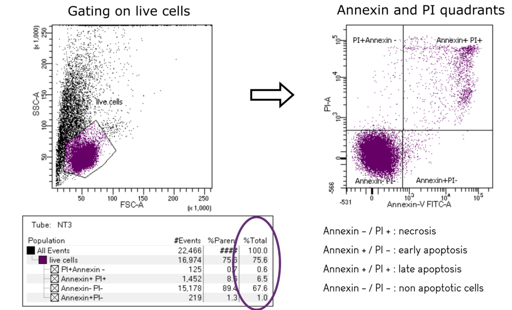
To better describe the activity of your agents we determine the apoptotic status of cells exposed to your compounds, using Annexin V/PI staining and by the analysis of caspase-3 and pro- or anti-apoptotic proteins (such as Bcl2 family proteins).

Antineo is able to complete the data obtained in end-point assays by continuous-monitoring assays (see continuous-monitoring).

Multiple colour flow cytometry

Multiple colour flow cytometry

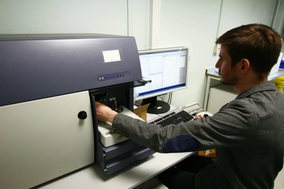
Antineo routinely performs flow cytometry studies (BD™ LSRFortessa and BD Canto™ flow cytometers, as well as CYTEK Aurora™ for spectral flow cytometry) for qualitative and quantitative multiparametric analyses, combined with FlowJo® LLC analyses.

We can combine up to 29 colours to explore in-depth your samples, or perform immunophenotyping studies.

Immunology and Immuno-oncology

Immunology and Immuno-oncology

Antineo’s panel of immunology assays are designed to help you understand the immunomodulatory effect of your compounds.

The list below is not exhaustive of what we can do, and as scientists, we are always eager to discuss with our clients and to design the experiment that will answer to their need.

T Cell based assays (human and murine cells):

  • T Cell activation assays characterise the activating potential of your compound compared with a standard anti-CD3/CD28 activation. We use either PBMCs or purified T Cells from a healthy donor. The readout of this assay is the production of IFNγ and TNFα by the activated T Cells. This can be quantified by flow cytometry or by ELISA.
  • T Cell proliferation assays measure the pro or anti-proliferative action of your compound by CFSE analysis by flow cytometry. Sub-set phenotyping of CD markers can be added to refine the analyses. This assay can be performed with continuous-monitoring.
  • Cytotoxic T Lymphocytes assays where activated T Cells are incubated with calcein-labelled tumour cells. The calcein release, measured by absorbance with a plate reader, is proportional to the cytotoxic properties of T Cells. This assay can be performed with continuous-monitoring.
  • Mixed Lymphocyte reactions identify agents modulating APC-mediated T Cell activation. The blood of two different healthy donors is used to co-culture T Cells and Dendritic Cells. The readout of T Cells activation and proliferation is the production of IFNγ and TNFα and incorporation of CSFE.

Myeloid cells based assays (human cells):

  • M1/M2 polarisation assays determine the capacity of your compound to reverse immune-suppressive signals. PBMCs or myeloid cells are differentiated in M1 or M2 macrophages.

Contact our Team to learn more

Contact