

AUTHORS
Marie TAUTOU, Charline PERROUIN, Jade RUARD, Léna MARLHOUX, Mélina GAUTHIER, Pierre-Antoine CHOFFOUR, Doriane MATHE, Charles DUMONTET
INTRODUCTION
This new luminescence-based method allows us to track tumors and metastases in in vivo models using our Ivis Imager®.
MATERIALS & METHODS
For the development of the luciferase bioluminescent cell lines tumor monitoring, we used SCID CB17 and NSG mice, with the MDA-MB-231 cell line, a model of breast cancer, and DEL cell line, a model of lymphoma.
Compared to the fluorescence method, luminescence is a more sensitive approach allowing whole body imaging and the detection of deep tumors and metastasis in live mice (1).
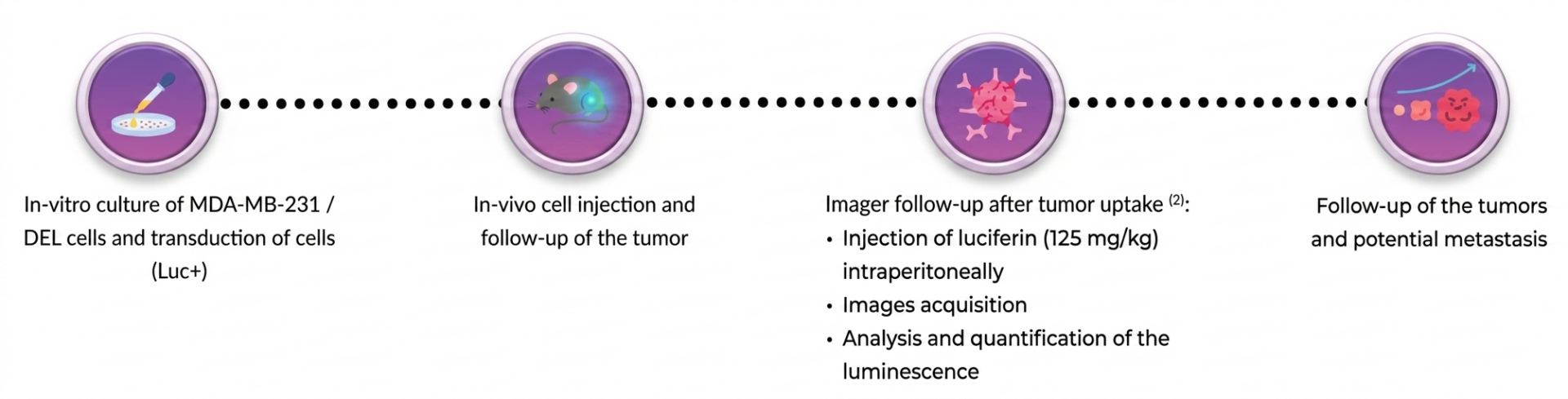
RESULTS
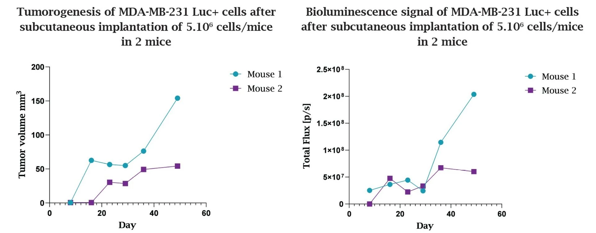
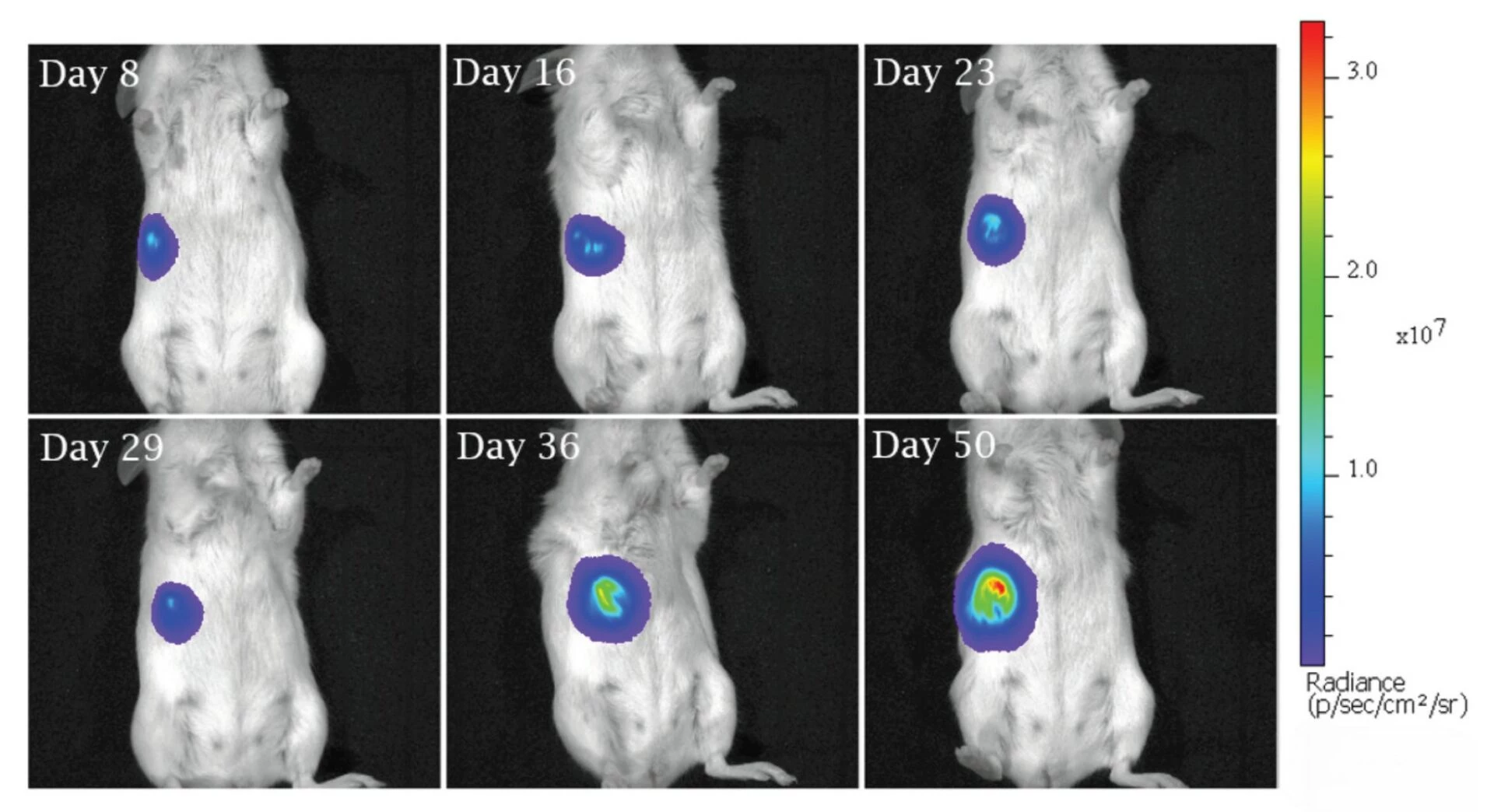
Monitoring of a breast cancer line – (MDA-MB-231 Luc+)
• Correlation between tumor volume measured with a caliper and the luciferase bioluminescent signal
• Consistent tumor growth after 2 months of monitoring
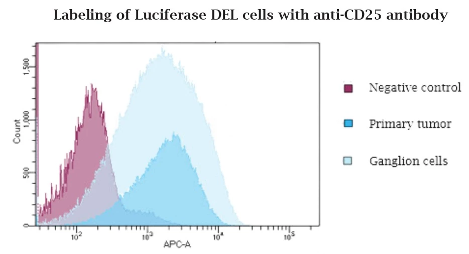
Monitoring of a metastasis from a T Lymphoma – (DEL Luc+)
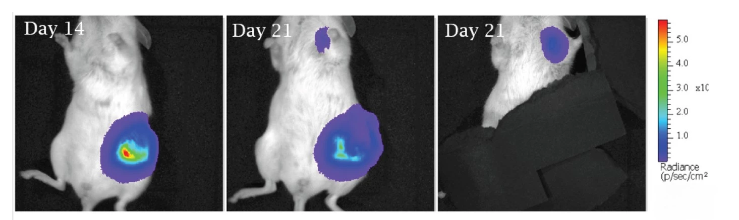
• Detection of a second signal different from the primary tumor, localized under the left paw
• Collection of the lymph node and dissociation of the cells for FACS analysis
• Validation of the second signal resulting of a metastasis from primary tumor
CONCLUSION
We have developed luciferase bioluminescent (Luc+) cell lines by transduction, and we have conducted a first longitudinal follow-up of the MDA-MB-231 cell line (breast cancer), and a second monitoring of the DEL cell line (lymphoma cancer), injected subcutaneously, and the associated metastasis.
The images obtained with the Ivis Imager® allow us to regularly monitor tumors and potential metastases over time and without loss of luminescence.
We also offer alternative experimental designs with the Ivis Imager®:
• Tracking of a luminescent compound to follow its biodistribution
• Orthotopic injection for a more precise and non-invasive follow-up with the classic method (caliper)
• Development of on-demand models
REFERENCES
1. Genevois, Coralie, et al. « In Vivo Follow-up of Brain Tumor Growth via Bioluminescence Imaging and Fluorescence Tomography ». International Journal of Molecular Sciences, vol. 17, no 11, octobre 2016, p. 1815.
2. Liu, Shirley, et al. « Brightening up Biology: Advances in Luciferase Systems for in Vivo Imaging ». ACS Chemical Biology, vol. 16, no 12, décembre 2021, p. 2707‑18.
FAQ
In preclinical oncology research, modified cell lineages engineered to express metabolic enzymes are utilized to track malignancy progression within living animal hosts. The processing of specialized light-emitting substrates by these modified lines generates measurable optical signals. These signals are routinely captured across whole organism bodies using advanced optical imaging systems like the Ivis Imager®. Research programs across academic entities and commercial organizations are supported through these specialized models. Evaluated options include both syngeneic models and xenograft preparations. In addition, standard care drug resistance patterns can be systematically assessed. Experimental protocols are carefully designed to match current scientific knowledge and experimental compounds.
Luminescence offers enhanced analytical sensitivity compared to traditional fluorescent imaging methods. Light signals generated by luciferase bioluminescent cells can penetrate deep internal tissue structures without the background interference often associated with external light excitation. Consequently, entire body scans are successfully performed on living subjects. Internal disease sites and secondary metastatic lesions are precisely localized through this approach. Non-invasive tracking allows repeated measurements to be gathered from the same host over time.
In vivo validation studies were conducted using immunocompromised mouse strains, specifically SCID CB17 and NSG mice. Two distinct cancer targets were selected to assess the system across different malignancy types. The MDA-MB-231 cell line was employed as a model for breast cancer. Lymphoma was investigated through the application of the DEL cell line. Both lineages were modified into luciferase bioluminescent versions to enable light emission upon substrate processing. Light patterns were subsequently tracked in real time across living murine hosts.
Engineered cellular lineages expressing functional enzymes are supplied with the substrate Luciferine. A chemical reaction occurs internally as the substrate is actively metabolized by modified luciferase bioluminescent cell strains. Visible optical light is emitted as a direct consequence of this enzymatic process. Emission levels correlate directly with the presence and activity of viable target cells within the subject. Signal intensity maps are then captured digitally using specialized detection instruments
Optical emissions generated by modified cellular lineages are recorded using the Ivis Imager® system. Whole body optical detection is facilitated by this specialized equipment. Live subjects are placed within the detection chamber following substrate administration. Accurate measurement of primary masses and distant secondary spreads is permitted by high-sensitivity sensors. Non-invasive monitoring is continuously maintained across longitudinal study timelines. Data acquired through this method support drug evaluation programs in oncology research.
Deeply seated internal metastases and primary masses are effectively detected using luciferase bioluminescent monitoring methods. High tissue penetration of emitted light signals allows clear visualization of internal sites. Secondary disease spread across various organs can be monitored in intact living subjects. Surgical intervention or subject termination is not required to observe internal structural changes.
Experimental designs are customized by aligning preclinical study parameters with current scientific literature and competing drug candidates. A variety of in vitro and in vivo testing strategies are offered to evaluate therapeutic efficacy. Selection of representative disease models includes both syngeneic host systems and human tissue xenografts. Modified luciferase bioluminescent cell lines are incorporated to enable precise quantitative tracking of treatment outcomes. Drug resistance characteristics are evaluated using tailored cell variants. Guidance is provided to accelerate research timelines for both corporate entities and academic research teams.
