Antineo provides a variety of services designed to accelerate the preclinical development of novel therapeutic antibodies. With our in vitro and ex vivo services, we highlight the target of your antibody, and decipher the mechanisms of action of your compounds. Our original in vivo experiments provide Proof of Concept strengthening your data.

Our offer includes

  • Immunology ex vivo assays (T cell activation, CTL assays, Macrophage polarisation etc.)
  • ADC characterization
  • Original in vitro and in vivo assays for bispecific antibodies (anti-CD3)
  • In vitro or ex vivo ADCC, ADCP and CDC assays
  • Original methods for in vivo assessment of ADCC and CDC activities
  • A unique panel of tumour models presenting secondary resistances to immunotherapies
Immunology and Immuno-oncology

Immunology and Immuno-oncology

Antineo’s panel of immunology assays are designed to help you understand the immunomodulatory effect of your compounds.

The list below is not exhaustive of what we can do, and as scientists, we are always eager to discuss with our clients and to design the experiment that will answer to their need.

T Cell based assays (human and murine cells):

  • T Cell activation assays characterise the activating potential of your compound compared with a standard anti-CD3/CD28 activation. We use either PBMCs or purified T Cells from a healthy donor. The readout of this assay is the production of IFNγ and TNFα by the activated T Cells. This can be quantified by flow cytometry or by ELISA.
  • T Cell proliferation assays measure the pro or anti-proliferative action of your compound by CFSE analysis by flow cytometry. Sub-set phenotyping of CD markers can be added to refine the analyses. This assay can be performed with continuous-monitoring.
  • Cytotoxic T Lymphocytes assays where activated T Cells are incubated with calcein-labelled tumour cells. The calcein release, measured by absorbance with a plate reader, is proportional to the cytotoxic properties of T Cells. This assay can be performed with continuous-monitoring.
  • Mixed Lymphocyte reactions identify agents modulating APC-mediated T Cell activation. The blood of two different healthy donors is used to co-culture T Cells and Dendritic Cells. The readout of T Cells activation and proliferation is the production of IFNγ and TNFα and incorporation of CSFE.

Myeloid cells based assays (human cells):

  • M1/M2 polarisation assays determine the capacity of your compound to reverse immune-suppressive signals. PBMCs or myeloid cells are differentiated in M1 or M2 macrophages.

ADC characterization

ADC characterization

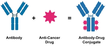
Antibody-drug conjugates (ADCs) are therapies combining the high activity of cytotoxic drugs with the high specificity of monoclonal antibodies to improve tumor targeting and reduce toxic side effects. ADCs are made of three main components : an antibody responsible for selectively recognizing cancer cell surface antigens, payloads responsible for killing cancer cells, and a linker to connect antibodies with the payloads.

Antineo can help you characterize your ADC with a complete set of in vitro and in vivo assays:

  • Cytotoxicity assays, Internalization assays
  • By-stander effect : Once treated by targeted cells, ADCs can release cytotoxic drug molecules that diffuse  into neighboring antigen-negative cells, thereby inducing their cytotoxicity. This effect can be assessed with a co-culture of two different cell lines and quantification based on fluorescence.
  • The antibody component of ADCs engage with immune effector cells to elicit antitumor immunity, including complement-dependent cytotoxicity (CDC), antibody-dependent cell cytotoxicity (ADCC), and antibody-dependent cell phagocytosis (ADCP) effects. Antineo proposes several assays to decipher such mechanisms both in vitro, ex/in vivo.
  • In vivo efficacy, volume-response
  • Lung toxicity : model of Intersticial lung disease (ILD)/pneumonitis, which is a significant adverse event related to some ADCs.
In vivo efficacy study for bispecific antibodies

In vivo efficacy study for bispecific antibodies

Bispecific antibodies can simultaneously bind two different types of antigen or two different cellular types : tumor cells and T-cells CD3, tumor cells and a radiolabeled hapten, or two different tumor cells antigens.

  • A screening of your bispecific antibodies can be performed to select the optimal clone : Binding assays, Quantification of antigen sites by Flow cytometry
  • Bispecifc antibodies cellular toxicity can be investigated
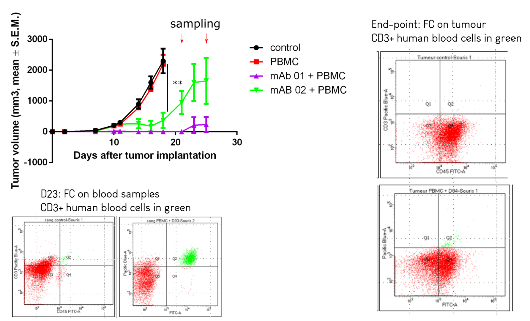
  • To assess the antitumor efficacy of bispecific antibodies targeting immune cells and tumour cells in vivo, we re-infuse human PBMC (Peripheral Blood Mononuclear Cells) or purified populations in immunodeficient mice implanted with a tumour model, and administer the treatment. Tumour growth is then evaluated in the different groups. The proliferation and activation status of human cells can be assessed at various time points by flow cytometry, and their presence in the tumour micro environment can be quantified at end points.


Antibody dependant mechanisms

Antibody dependant mechanisms

Some therapeutic monoclonal antibodies are able to recruit innate immune cells, or complement for destruction of tumour cells. Antineo proposes several assays to decipher such mechanisms both in vitro, ex/in vivo.

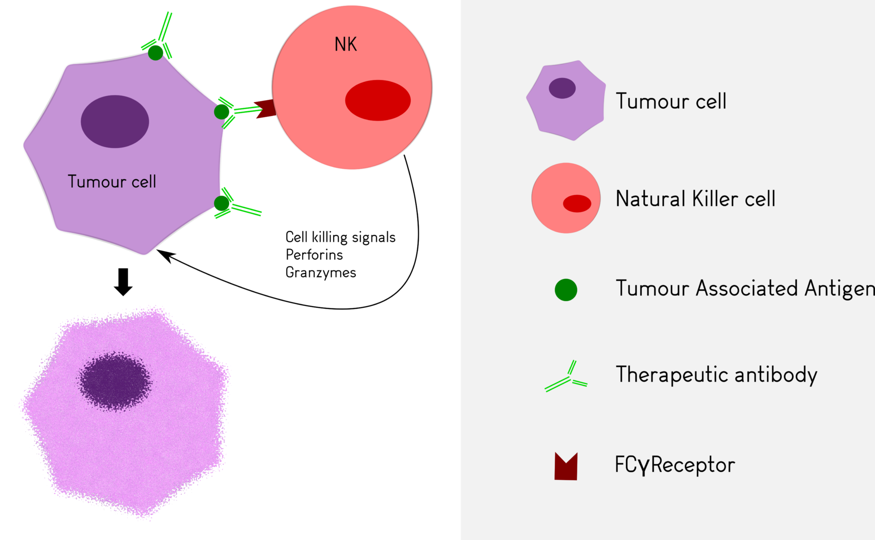
Antibody Dependent Cellular Cytotoxicity (ADCC)

The best described mechanisms involve Antibody Dependent Cellular Cytotoxicity in which cells, such as Natural Killer lymphocytes, bind the antibody through their Fcγ receptor and are activated to release cytotoxic products.
Antibody Dependent Cellular Phagocytosis (ADCP)

In Antibody Dependent Cellular Phagocytosis, phagocytic cells such as granulocytes or macrophages are engaged by the antibody via their Fcγ receptor to destroy tumour cells by phagocytosis.
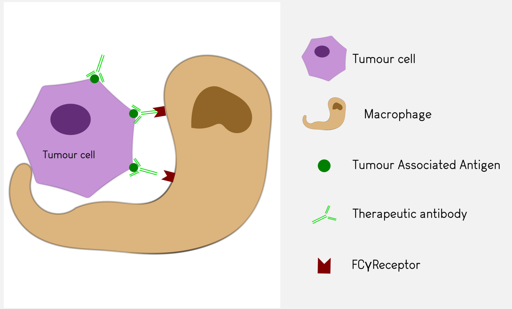
Antineo analyses the ability of your candidate antibodies to perform ADCC and/or ADCP using model cell lines or fresh human effector cells from healthy donors. ADCC is quantified using the release of calcein by tumour cells while ADCP is quantified by flow cytometry, evaluating the transfer of tumour cell fluorescence to effector cells. Both assays can be can be performed with continuous-monitoring, and ADCC can be assessed with our original in vivo assay.

Complement-Dependent Cytotoxicity (CDC)

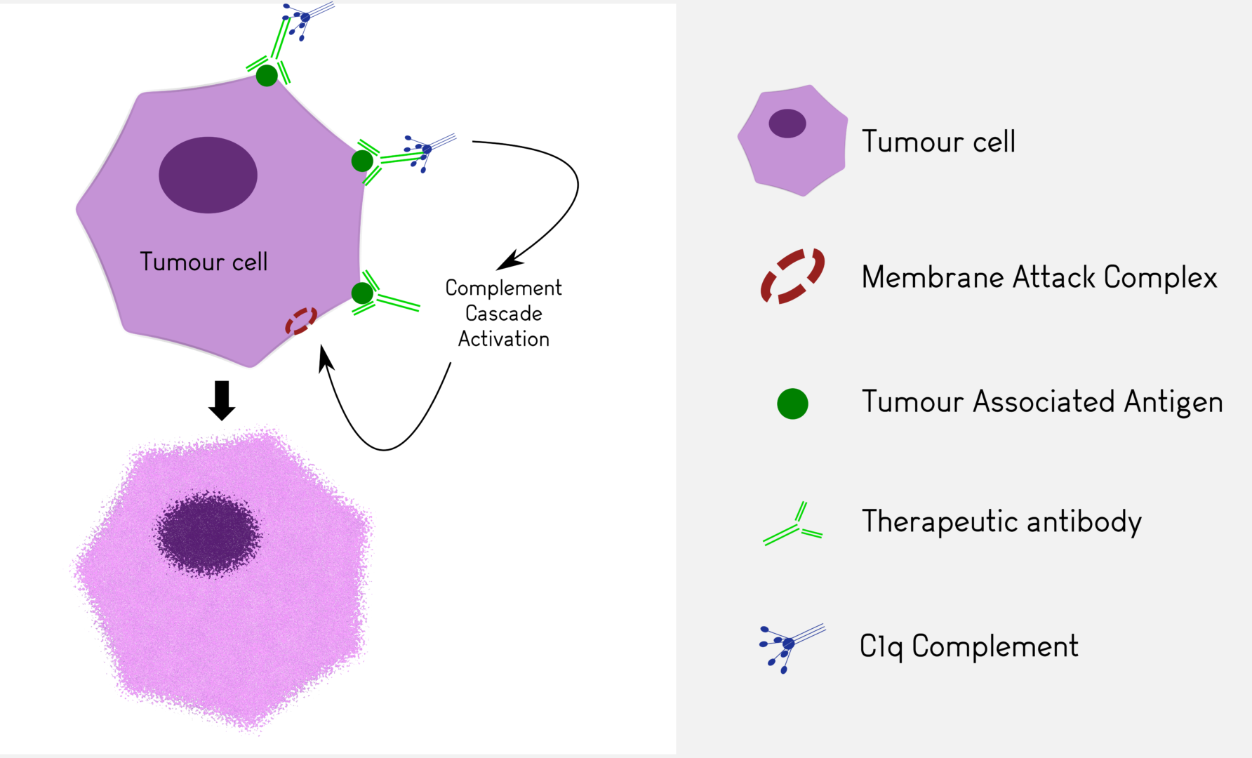
We determine the effect of your antibody on cell lines or fresh human samples in presence of human or rabbit serum containing complement. CDC assays are performed using calcein release. These tests can be completed in vivo using cobra venom as a complement depleting agent (in vivo studies).

In vivo ADCC assay

In vivo ADCC assay

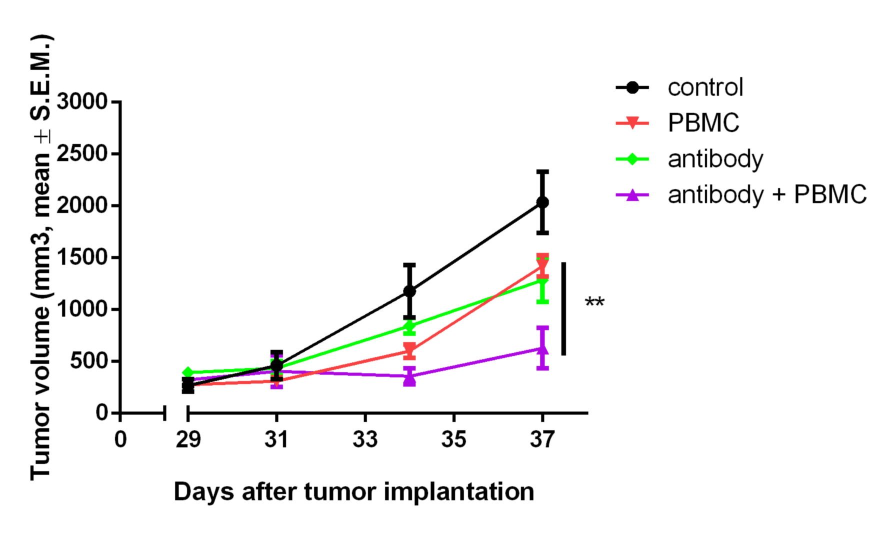
We perform in vivo ADCC (Antibody Dependent Cell Cytotoxicity) studies using cell lines and purified normal blood cell fractions. These may consist of total human leucocytes (Peripheral Blood Mononuclear Cells) from healthy donors or purified subpopulations (granulocytes, NK cells, T cells, monocytes). When the therapeutic antibody possesses ADCC activity, the antitumor efficacy of the compound in presence of blood cells is increased.

In vivo CDC assay

In vivo CDC assay

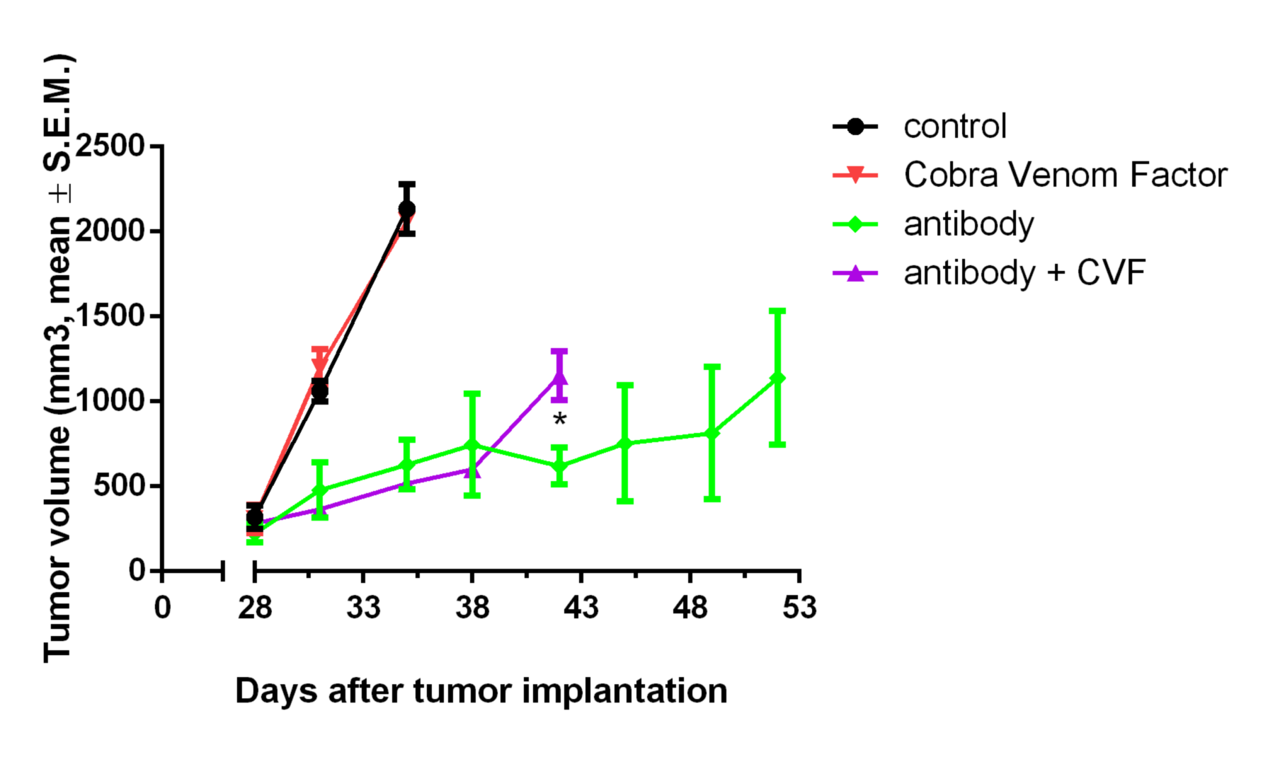
The CDC component is evaluated in vivo by administration of cobra venom factor, an activator of the complement system used to perform in vivo depletion.

Our protocol consists in the injection/implantation of a tumour model and administration of the therapeutic antibody in the prophylactic or therapeutic setting, in the presence or absence of cobra venom. When CDC is a key component of the antitumor activity of an antibody, the effect of the antibody on tumour growth will be reduced by prior administration of cobra venom factor.

Contact our Team to learn more

Contact