Cell Culture

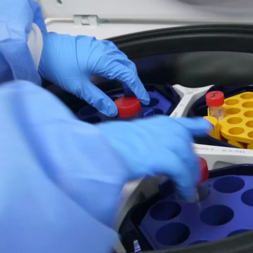
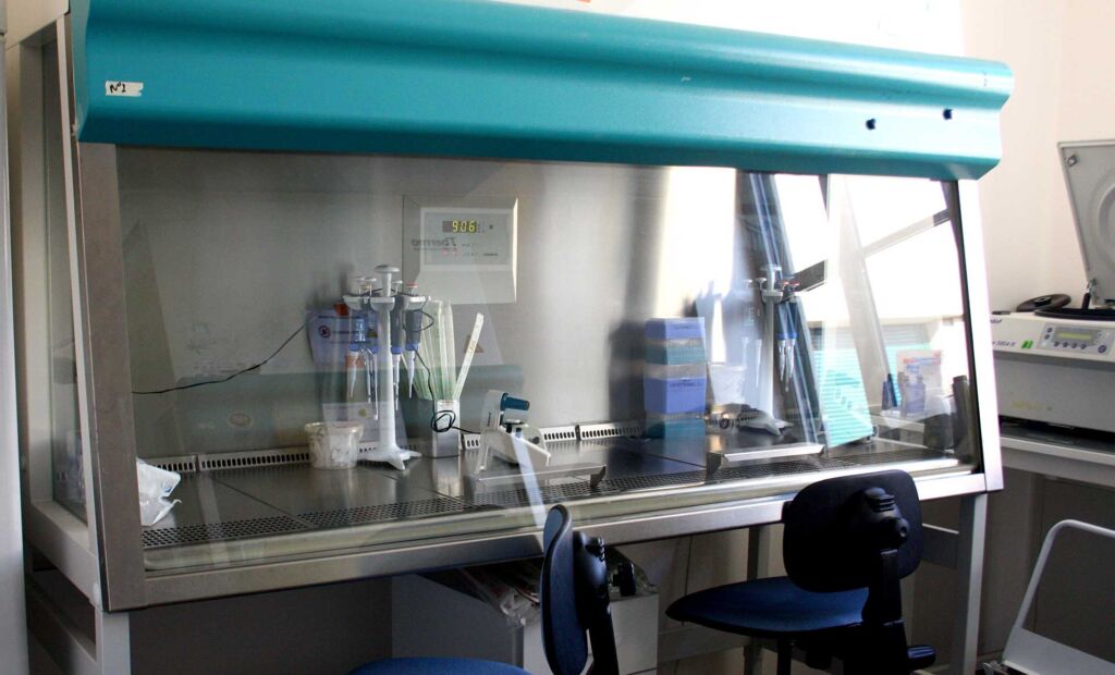
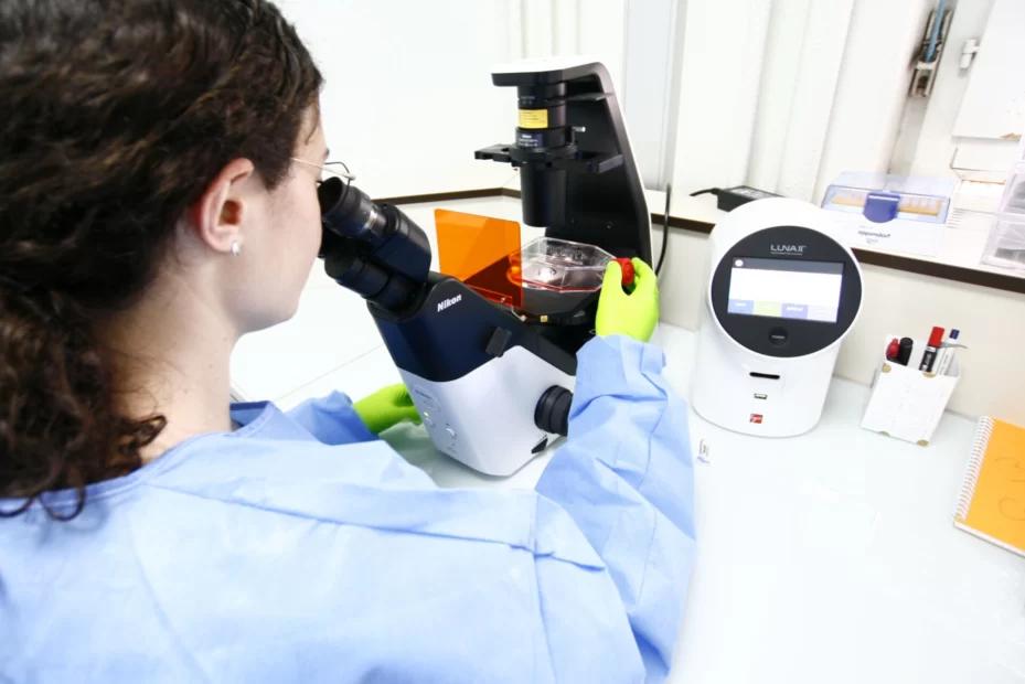
Our cell culture facility is maintained in a controlled atmosphere environment that meets P2 microbiological security standards. This dedicated space is designed to ensure the necessary quality and reliability to work with both human and murine cells. Featuring cell counters, inverted contrast phase microscopy, and fluorescent microscopes, our facility is fully equipped to support cell culture work under both normoxic and hypoxic conditions.

For example, this setup is ideal to study cellular responses to oxygen levels variation, which is particularly important in cancer research where the tumor microenvironment can have an impact on cellular behavior and therapeutic responses.

xCELLigence® and Incucyte® technologies enable continuous monitoring of cell proliferation, in real-time. With xCELLigence® we can track cellular activities such as proliferation, adhesion, and migration. The Incucyte® allows us to monitor cell growth, morphologic changes, and even cell death, making it a powerful tool for evaluating the effects of various treatments, such as cytotoxic drugs or gene therapies, on cell viability and function.


 

Flow Cytometry

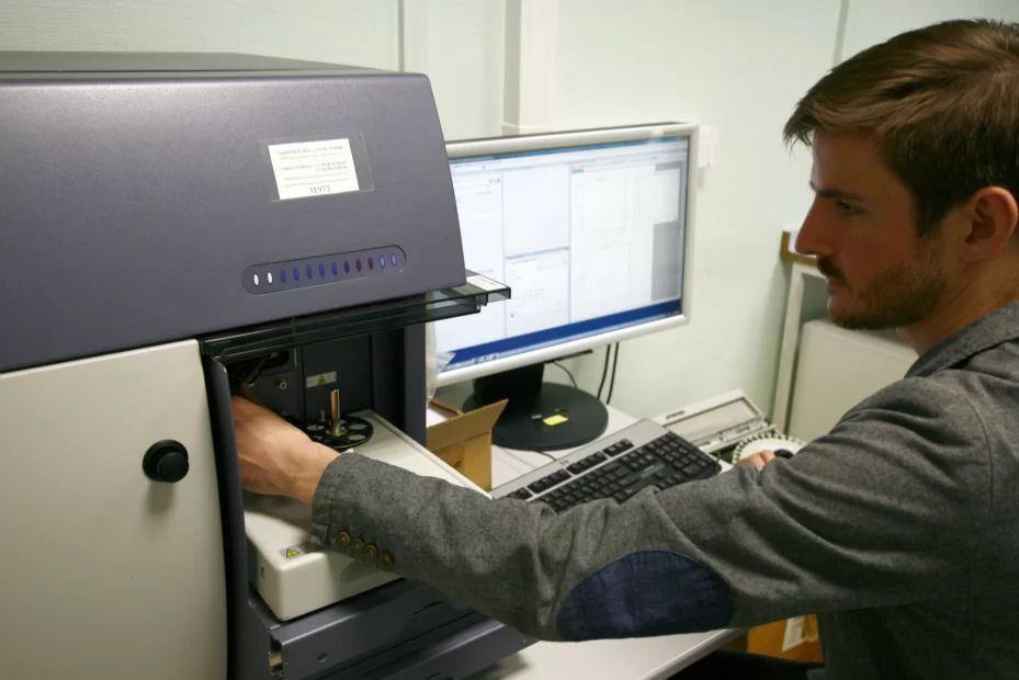
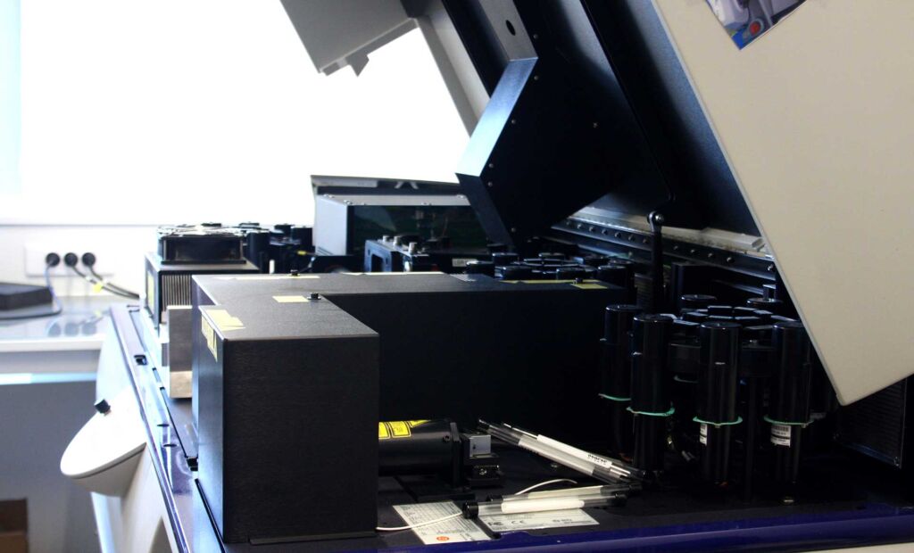
Flow cytometry is a useful tool for a wide range of applications, including immunophenotyping of treated tumors and cytokine profiling. We routinely perform flow cytometry studies to provide in-depth, multiparametric analyses of our clients’ samples. Our flow cytometry capabilities include the BD™ Quanto and LSRFortessa™ flow cytometers, as well as the spectral analyzer CYTEK Aurora™ for qualitative and quantitative analysis of cell populations. These systems allow us to detect and quantify various markers on the surface and inside of cells, providing detailed insights into cellular profiles and responses to treatments.

  • Multiparametric analysis :

With the ability to analyze up to 29 parameters simultaneously, our flow cytometers allow highly detailed profiling of cell populations, enabling the identification of subtle variations in subpopulations.

  • Tailored antibody panels:

Our scientific team has the expertise to build fully customized flow cytometry panels, depending on what you want to investigate in your samples.

  • Data analysis 

We use FlowJo® LLC software to analyze and interpret flow cytometry data, enabling gating, clustering, and visualization of complex data sets. This enables us to provide detailed, actionable information on cell function, phenotype, and response to treatment.


Small animal Imaging

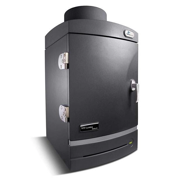
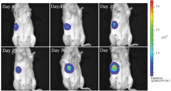
We use our Luciferase (Luc+) cell lines together with the Ivis Lumina Imager® (Perkin-Elmer) as part of our imaging platform. For high-sensitivity luminescence-based imaging studies, these cell lines are designed to express luciferase, an enzyme that breaks down luciferin and produces light that the Ivis Lumina Imager® can detect. Our small animal imaging capabilities allow for real-time, non-invasive tumor and metastasis monitoring in in vivo models.

  • Longitudinal tumor monitoring in efficacy studies:

Key information on tumor growth and progression can be obtained by using luminescence to track tumors and metastases in live mice throughout time with greater sensitivity and accuracy. Whole-body imaging is made possible by this technology, which makes it possible to follow tumors and metastases.

  • Tracking luminescent compounds for biodistribution studies:

By monitoring the biodistribution of luminous substances, we may assess how therapies are absorbed by the body and how they accumulate in tumors or other tissues.

  • Orthotopic tumor models:

Orthotopic implantations, in which tumors are placed directly into the tissue from which they originated (such as the breast, liver, or lung), are an option we provide. Compared to conventional techniques (such as caliper measurement), our approach provides a more accurate, physiological tumor model that replicates the real tumor microenvironment and permits more precise tracking as tumor growth and treatment efficacy assessment.

  • On-demand model development:

We can also develop customized, on-demand models for specific research needs, whether for novel tumor types, rare cancer models, or therapeutic testing in a particular model.

These imaging capabilities provide critical, non-invasive insights into tumor biology, therapeutic efficacy, and the dynamics of cancer progression, all of which are essential for developing and evaluating novel cancer therapies.


3D Bioprinting

3D Bioprinting with BIOX® from Cellink

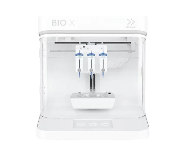
We are also equipped with state-of-the-art 3D bioprinting technology thanks to the BIOX® printer from Cellink. This advanced technology enables us to generate 3D tissue models, to reproduce the human cancer microenvironment more accurately than conventional 2D cultures.

With its unique ability to print various bioinks at the same time, such as human cells, tumor cell lines, and extracellular matrix components, the BIOX® 3D bioprinter enables the design of customized 3D structures that reflect the true complexity and heterogeneity of tumors. This technology enables the creation of more realistic tumor models for drug screening, facilitating the models' choice for in vivo experiments afterward.

BIOX® also makes it possible to integrate various cells into the model, such as immune cells, stromal cells, and endothelial cells, to analyze cell-cell interactions and simulate the tumor microenvironment. This method is valuable for the evaluation of new treatments, such as targeted drugs or immunotherapies, in addition to the possibility of designing personalized models based on patients' tumor cells.

Thanks to the precision and flexibility of the BIOX® printer, we are able to create customized 3D models suitable for a variety of testing applications, such as drug efficacy monitoring, toxicity monitoring, and tumor invasion studies. By using 3D-printed tissue models, we are able to offer more reliable and reproducible results, overcoming the limitations of preclinical studies and accelerating the preclinical development of anti-cancer therapeutics.

Contact our Team to learn more

Contact